

Platelet count reduction during in vitro membrane oxygenation affects platelet activation, neutrophil extracellular trap formation and clot stability, but does not prevent clotting
- PMID: 33475044
- PMCID: PMC8928426
- DOI: 10.1177/0267659121989231
Platelet count reduction during in vitro membrane oxygenation affects platelet activation, neutrophil extracellular trap formation and clot stability, but does not prevent clotting
Abstract
Introduction: Due to improved technology and increased application the mortality during extracorporeal membrane oxygenation (ECMO) is constantly declining. Nevertheless, complications including haemorrhage or thrombus formation remain frequent. Local mitigation of coagulation within an ECMO system to prevent thrombus formation on ECMO components and optimizing systemic anticoagulation is an approach to reduce clotting and bleeding complications at once. Foreign surfaces of ECMO systems, activate platelets (PLTs), which besides their major role in coagulation, can trigger the formation of neutrophil extracellular traps (NETs) contributing to robust thrombus formation. The impact of a reduced PLT count on PLT activation and NET formation is of paramount importance and worth investigating.
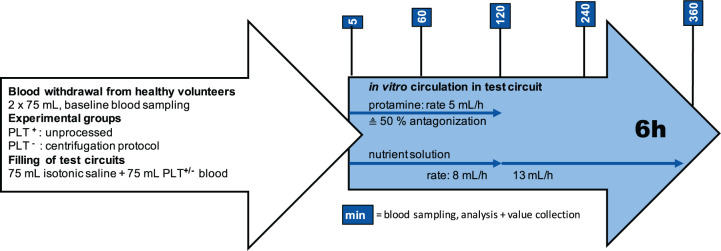
Methods: In this study platelet poor (PLT-) and native (PLT+) heparinized human blood was circulated in two identical in vitro test circuits for ECMO devices for 6 hours. PLT reduction was achieved by a centrifugation protocol prior to the experiments. To achieve native coagulation characteristics within the test circuits, the initial heparin dose was antagonized by continuous protamine administration.
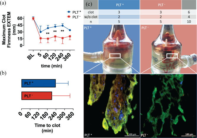
Results: The PLT- group showed significantly lower platelet activation, basal NET formation and limited clot stability measured via thromboelastometry. Fluorescent and scanning electron microscope imaging showed differences in clot composition. Both groups showed equal clot formation within the circuit.
Conclusions: This study demonstrated that the reduction of PLTs within an ECMO system is associated with limited PLT activation and NET formation, which reduces clot stability but is not sufficient to inhibit clot formation per se.
Keywords: clot stability; extracorporeal membrane oxygenation (ECMO); in vitro test circuit; neutrophil extracellular trap (NET) formation; platelet activation.
Conflict of interest statement
Figures


References
-
- Brodie D, Bacchetta M. Extracorporeal membrane oxygenation for ARDS in adults. N Engl J Med 2011; 365: 1905–1914. - PubMed
-
- Munshi L, Walkey A, Goligher E, et al. Venovenous extracorporeal membrane oxygenation for acute respiratory distress syndrome: a systematic review and meta-analysis. Lancet Respir Med 2019; 7: 163–172. - PubMed
-
- Zapol WM, Snider MT, Hill JD, et al. Extracorporeal membrane oxygenation in severe acute respiratory failure. A randomized prospective study. JAMA 1979; 242: 2193–2196. - PubMed
-
- Combes A, Hajage D, Capellier G, et al. Extracorporeal membrane oxygenation for severe acute respiratory distress syndrome. N Engl J Med 2018; 378: 1965–1975. - PubMed
MeSH terms
LinkOut - more resources
Full Text Sources
Other Literature Sources
Medical
