

Parkinson-like early autonomic dysfunction induced by vagal application of DOPAL in rats
- PMID: 33475253
- PMCID: PMC8025611
- DOI: 10.1111/cns.13589
Parkinson-like early autonomic dysfunction induced by vagal application of DOPAL in rats
Abstract
Aim: To understand why autonomic failures, a common non-motor symptom of Parkinson's disease (PD), occur earlier than typical motor disorders.
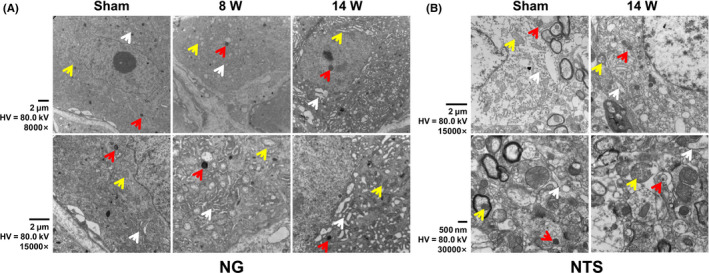
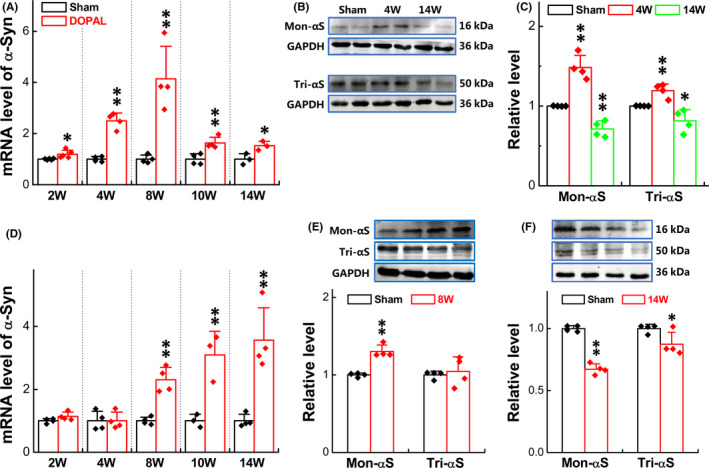
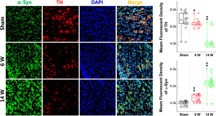
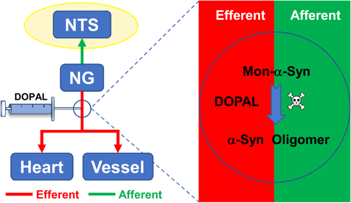
Methods: Vagal application of DOPAL (3,4-dihydroxyphenylacetaldehyde) to simulate PD-like autonomic dysfunction and understand the connection between PD and cardiovascular dysfunction. Molecular and morphological approaches were employed to test the time-dependent alternation of α-synuclein aggregation and the ultrastructure changes in the heart and nodose (NG)/nucleus tractus solitarius (NTS).
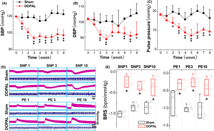
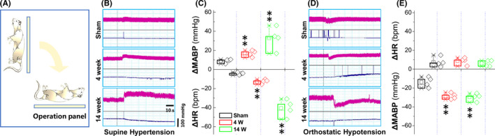
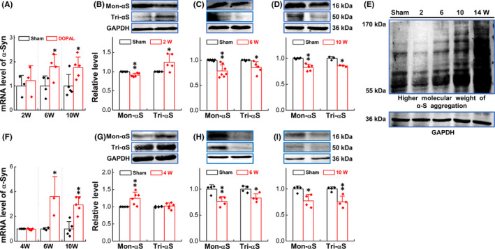
Results: Blood pressure (BP) and baroreflex sensitivity of DOPAL-treated rats were significantly reduced accompanied with a time-dependent change in orthostatic BP, consistent with altered echocardiography and cardiomyocyte mitochondrial ultrastructure. Notably, time-dependent and collaborated changes in Mon-/Tri-α-synuclein were paralleled with morphological alternation in the NG and NTS.
Conclusion: These all demonstrate that early autonomic dysfunction mediated by vagal application of DOPAL highly suggests the plausible etiology of PD initiated from peripheral, rather than central site. It will provide a scientific basis for the prevention and early diagnosis of PD.
Keywords: DOPAL; Parkinson's disease; autonomic dysfunction; vagus; α-synuclein.
© 2021 The Authors. CNS Neuroscience & Therapeutics Published by John Wiley & Sons Ltd.
Conflict of interest statement
The authors declare that there is no conflict of interest associated with the contents of this article.
Figures

Similar articles
-
Renalase Overexpression-Mediated Excessive Metabolism of Peripheral Dopamine, DOPAL Accumulation, and α-Synuclein Aggregation in Baroreflex Afferents Contribute to Neuronal Degeneration and Autonomic Dysfunction.Biomedicines. 2025 May 20;13(5):1243. doi: 10.3390/biomedicines13051243. Biomedicines. 2025. PMID: 40427069 Free PMC article.
-
Role of Parkinson's Disease-Linked Mutations and N-Terminal Acetylation on the Oligomerization of α-Synuclein Induced by 3,4-Dihydroxyphenylacetaldehyde.ACS Chem Neurosci. 2019 Jan 16;10(1):690-703. doi: 10.1021/acschemneuro.8b00498. Epub 2018 Nov 5. ACS Chem Neurosci. 2019. PMID: 30352158 Free PMC article.
-
Aldehyde adducts inhibit 3,4-dihydroxyphenylacetaldehyde-induced α-synuclein aggregation and toxicity: Implication for Parkinson neuroprotective therapy.Eur J Pharmacol. 2019 Feb 15;845:65-73. doi: 10.1016/j.ejphar.2018.12.027. Epub 2018 Dec 21. Eur J Pharmacol. 2019. PMID: 30579934
-
3,4-dihydroxyphenylacetaldehyde: a potential target for neuroprotective therapy in Parkinson's disease.Curr Drug Targets CNS Neurol Disord. 2003 Apr;2(2):143-8. doi: 10.2174/1568007033482913. Curr Drug Targets CNS Neurol Disord. 2003. PMID: 12769806 Review.
-
Cardiovascular autonomic dysfunction in animal models of Parkinson's disease.J Parkinsons Dis. 2011;1(4):321-7. doi: 10.3233/JPD-2011-11042. J Parkinsons Dis. 2011. PMID: 23933655 Review.
Cited by
-
The Catecholaldehyde Hypothesis for the Pathogenesis of Catecholaminergic Neurodegeneration: What We Know and What We Do Not Know.Int J Mol Sci. 2021 Jun 1;22(11):5999. doi: 10.3390/ijms22115999. Int J Mol Sci. 2021. PMID: 34206133 Free PMC article. Review.
-
Synchronous monitoring of brain-heart electrophysiology using heart rate variability coupled with rapid quantitative electroencephalography in orthostatic hypotension patients with α-synucleinopathies: Rapid prediction of orthostatic hypotension and preliminary exploration of brain stimulation therapy.CNS Neurosci Ther. 2024 Feb;30(2):e14571. doi: 10.1111/cns.14571. CNS Neurosci Ther. 2024. PMID: 38421092 Free PMC article.
-
Estrogen-dependent depressor response of melatonin via baroreflex afferent function and intensification of PKC-mediated Nav1.9 activation.Acta Pharmacol Sin. 2022 Sep;43(9):2313-2324. doi: 10.1038/s41401-022-00867-w. Epub 2022 Feb 7. Acta Pharmacol Sin. 2022. PMID: 35132193 Free PMC article.
-
Oxidative Transformations of 3,4-Dihydroxyphenylacetaldehyde Generate Potential Reactive Intermediates as Causative Agents for Its Neurotoxicity.Int J Mol Sci. 2021 Oct 29;22(21):11751. doi: 10.3390/ijms222111751. Int J Mol Sci. 2021. PMID: 34769179 Free PMC article.
-
Disruption of Dopamine Homeostasis Associated with Alteration of Proteins in Synaptic Vesicles: A Putative Central Mechanism of Parkinson's Disease Pathogenesis.Aging Dis. 2024 May 7;15(3):1204-1226. doi: 10.14336/AD.2023.0821-2. Aging Dis. 2024. PMID: 37815908 Free PMC article. Review.
References
-
- Schapira AHV, Chaudhuri KR, Jenner P. Non‐motor features of Parkinson disease. Nat Rev Neurosci. 2017;18:509. - PubMed
-
- Kuhlenbaumer G, Berg D. Parkinson disease genetics: too early to predict progression? Nat Rev Neurol. 2019;15:625‐626. - PubMed
-
- Sartori M, Pessina AC. Orthostatic hypotension and supine hypertension in pure autonomic failure. Ital HeartJ Suppl. 2004;5:879‐882. - PubMed
-
- Espay AJ, LeWitt PA, Hauser RA, et al. Neurogenic orthostatic hypotension and supine hypertension in Parkinson's disease and related synucleinopathies: prioritisation of treatment targets. Lancet Neurol. 2016;15:954‐966. - PubMed
Publication types
MeSH terms
Substances
LinkOut - more resources
Full Text Sources
Other Literature Sources
Medical
