. 2021 Jan 1;38(1):1-29.
doi: 10.1097/WNP.0000000000000806.

American Clinical Neurophysiology Society's Standardized Critical Care EEG Terminology: 2021 Version

Affiliations

American Clinical Neurophysiology Society's Standardized Critical Care EEG Terminology: 2021 Version

Lawrence J Hirsch et al. J Clin Neurophysiol. .
No abstract available


Conflict of interest statement

L. J. Hirsch received consultation fees from Aquestive, Ceribell, Marinus, Medtronic, Neuropace and UCB; received authorship royalties from Wolters Kluwer and Wiley; and received honoraria for speaking from Neuropace and Natus. S. M. LaRoche received royalties from Demos/Springer Publishing. S. Beniczky is consultant for Brain Sentinel & Epihunter and Philips; speaker for Eisai, UCB, GW Pharma, Natus, BIAL; and received research grants from Brain Sentinel, Philips, Eisai, UCB, GW Pharma, Natus, BIAL, Epihunter, Eurostars (EU), Independent Research Fund Denmark, Filadelfia Research Foundation, Juhl Foundation, Hansen Foundation. N. S. Abend received royalties from Demos; grants from PCORI and Epilepsy Foundation; and an institutional grant from UCB Pharma. J. W. Lee received grants from Bioserenity, Teladoc, Epilepsy Foundation; is co-founder of Soterya Inc; is a board member of the American Clinical Neurophysiology Society; does consulting for Biogen; and is site PI for Engage Therapeutics and NIH/NINDS R01-NS062092. C. J. Wustof does consulting for Persyst and PRA Health Care. C. D. Hahn received grants from Takeda Pharmaceuticals, UCB Pharma, Greenwich Biosciences. M. B. Westover is co-founder of Beacon Biosignals. E. E. Gerard received grants from Greenwich Pharmaceuticals, Xenon Pharmaceuticals, Sunovion, and Sage. S. T. Herman received grants from UCB Pharma, Neuropace, Sage. H. A. Haider receives author royalties from UpToDate and Springer; does consulting for Ceribell, and is on advisory board for Eisai. A. Rodriguez-Ruiz is co-owner of Rodzi LLC which has no relationship to this work. E. J. Gilmore received a grant from UCB Pharma. J. Claassen is a shareholder of iCE Neurosystems and received a grant from McDonnell Foundation. A, M. Husain received grants from UCB Pharma, Jazz Pharma, Biogen Idec; and received payment from Marinus Pharma, Eisai Pharma, Neurelis Pharma, Blackthorn Pharma, Demos/Springer and Wolters Kluwer publishers. J. Y. Yoo received grants from NIH NeuroNEXT, Zimmer Biomet, LVIS; and receives author royalties from Elsevier. P. W. Kaplan receives author royalties from Demos and Wiley publishers; does consulting for Ceribell; and is expert witness qEEG. M. R. Nuwer is a shareholder of Corticare. M. van Putten is co-founder of Clinical Science Systems. R. Sutter received grants from Swiss National Foundation (No 320030_169379), and UCB Pharma. F. W. Drislane received a grant from American Academy of Neurology. E. Trinka discloses fees received from UCB, Eisai, Bial, Böhringer Ingelheim,Medtronic, Everpharma, GSK, Biogen, Takeda, Liva-Nova, Newbridge, Novartis, Sanofi, Sandoz, Sunovion, GW Pharmaceuticals, Marinus, Arvelle; grants from Austrian Science Fund (FWF), Österreichische Nationalbank, European Union, GSK, Biogen, Eisai, Novartis, Red Bull, Bayer, and UCB; other from Neuroconsult Ges.m.b.H., has been a trial investigator for Eisai, UCB, GSK, Pfitzer. The remaining authors have no funding or conflicts of interest to disclose.

Figures

FIG. 1.
FIG. 1.
A. Symmetric vs mild asymmetry in voltage. B. Symmetric vs mild asymmetry in frequency. C. Marked asymmetry in voltage and frequency.
FIG. 2.
FIG. 2.
Continuity. Percentages for each category refer to the percentage of the record that is “attenuated” or “suppressed.” How this percentage is derived is demonstrated in Fig. 4, page 6.
FIG. 3.
FIG. 3.
Discharge vs. Burst. *Phase: an area under the curve on one side of the baseline (see Section C 3d, page 13, and Fig. 23, page 13).
FIG. 4.
FIG. 4.
Attenuation percent or Suppression percent: the percent of the record/epoch that is attenuated or suppressed. This can range from 1% to 99%. If <1%, it is considered continuous. If >99%, it is considered either suppressed or attenuated, but not discontinuous. For example, a record with 2 second bursts alternating with 8 seconds of suppression, as shown here, would be Burst-Suppression with a suppression percent of 80%.
FIG. 5.
FIG. 5.
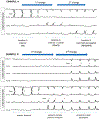
Localization of bursts. A. Generalized bursts, shifting predominance based on asynchrony. Symmetric bursts, at times starting on the left and others on the right, but never consistently the same side. This would be an example of generalized bursts, with shifting predominance based on asynchrony (rather than asymmetry, where they would sometimes be of greater amplitude on the left and other times the right). B. Lateralized bursts, bilateral asynchronous. Symmetric bursts consistently starting on the left with a lag before being seen on the right. This is an example of lateralized, bilateral asynchronous bursts. They are not Bilateral Independent (BI) bursts because there is a consistent relationship between the activity between hemispheres, i.e. the patterns are not independent.
FIG. 6.
FIG. 6.
A. Highly Epileptiform Bursts. --- dashed lines represent longer duration of suppression; ED epileptiform discharge. B. Highly Epileptiform Bursts. --- dashed lines represent longer duration of suppression.
FIG. 7.
FIG. 7.
A. Identical Bursts. The first 0.5 seconds or longer of each burst are visually similar in all channels (though only 1 channel shown) in most (>90%) bursts. B. Identical Bursts in a Stereotyped Cluster. The first 0.5 seconds or longer of each of 2 or more bursts in a stereotyped cluster are visually similar in all channels (though only 1 channel shown) in most (>90%) bursts.
FIG. 8.
FIG. 8.
State changes. At least 2 sustained types of background EEG, where: 1. The background activity is related to level of alertness or stimulation. 2. Each must persist ≥60 seconds to qualify as a “state”. 3. Stimulation should be able to transition the patient from the less alert to more alert/more stimulated state. 4. The more alert/more stimulated state is considered the “reported background” EEG. 5. State changes can also occur spontaneously. STIM = stimulation, Spont. = spontaneous. EEG background 1: stimulated/more awake: used for background feature description (“reported background”) EEG background 2: unstimulated/less awake state; commonly lasts minutes to hours (minimum: 60 s)
FIG. 9.
FIG. 9.
Cyclic Alternating Pattern of Encephalopathy (CAPE). Changes in EEG background between pattern 1 and pattern 2, where: 1. Each pattern lasts at least 10 seconds, 2. Spontaneously alternates between the two patterns in a regular manner, 3. For at least 6 cycles.
FIG. 10.
FIG. 10.
Anterior-posterior (AP) gradient.
FIG. 11.
FIG. 11.
Sporadic Epileptiform Discharges.
FIG. 12.
FIG. 12.
Polyspike versus BIRDs versus Highly Epileptiform Bursts.
FIG. 13.
FIG. 13.
Generalized Periodic Discharges (GPDs). Generalized: Bilateral synchronous and symmetric periodic discharges. In this case, the pattern is “frontally predominant.”
FIG. 14.
FIG. 14.
Lateralized Periodic Discharges (LPDs, unilateral). Unilateral: Periodic discharges only seen in one hemisphere (in this case left).
FIG. 15.
FIG. 15.
Lateralized Periodic Discharges (LPDs, bilateral asymmetric). Bilateral asymmetric: Periodic discharges seen bilaterally but clearly and consistently (>80% of the time) higher amplitude over one hemisphere (in this case left).
FIG. 16.
FIG. 16.
Lateralized Periodic Discharges (LPDs, bilateral asynchronous). Bilateral asynchronous: Periodic discharges seen bilaterally but clearly and consistently (>80% of the time) earlier on one side (in this case left). These are not Bilateral Independent (BI) because the latency between hemispheres is fixed (i.e., they are not independent populations).
FIG. 17.
FIG. 17.
Bilateral Independent Periodic Discharges (BIPDs). In BIPDs, lateralized patterns occur in each hemisphere asynchronously and at different frequencies.
FIG. 18.
FIG. 18.
Unilateral Independent Periodic Discharges (UIPDs). In UIPDs, periodic discharges occur in two independent locations simultaneously with both populations within a single hemisphere (in this case left).
FIG. 19.
FIG. 19.
Multifocal Periodic Discharges (MfPDs). In MfPDs, periodic discharges occur in three independent locations simultaneously with at least one in each hemisphere. If all three populations occurred within a single hemisphere this would remain UIPDs.
FIG. 20.
FIG. 20.
Periodic Discharges (PDs). 1. Repetition of a waveform with relatively uniform morphology and duration, 2. with a clearly discernable interdischarge interval between consecutive waveforms, and 3. recurrence of the waveform at nearly regular intervals: having a cycle length (i.e., period) varying by <50% from one cycle to the next in the majority (>50%) of cycle pairs. A pattern can qualify as rhythmic or periodic if and only if it continues for at least 6 cycles (e.g. 1 Hz for 6 seconds, or 3 Hz for 2 seconds).
FIG. 21.
FIG. 21.
Rhythmic Delta Activity (RDA). 1. Repetition of a waveform with relatively uniform morphology and duration and 2. without an interval between consecutive waveforms. 3. The duration of one cycle (i.e., the period) of the rhythmic pattern should vary by <50% from the duration of the subsequent cycle for the majority (>50%) of cycle pairs to qualify as rhythmic. A pattern can qualify as rhythmic or periodic if and only if it continues for at least 6 cycles (e.g. 1 Hz for 6 seconds, or 3 Hz for 2 seconds).
FIG. 22.
FIG. 22.
“Spike-and-wave” or “Sharp-and-wave” (SW). Spike-and-wave or Sharp-and-wave (SW): Polyspike, spike, or sharp wave consistently followed by a slow wave in a regularly repeating and alternating pattern (spike-wave-spike-wave-spike-wave), with a consistent relationship between the spike (or polyspike or sharp wave) component and the slow wave for at least 6 cycles; and with no interval between one spike-wave complex and the next (if there is an interval, this would qualify as PDs, where each discharge is a spike-and-wave).
FIG. 23.
FIG. 23.
The Number of Phases. Number of Phases = 1 + number of baseline crossings of the typical discharge. In this case there are a total of 2 baseline crossings, therefore the number of phases is 1 + 2 = 3 phases. A phase is the part of the signal above or below the imaginary baseline. In this case, phase 1 (pink) is above, phase 2 (blue) is below, and phase 3 (yellow) is above again.
FIG. 24.
FIG. 24.
Evolution of frequency. At least 2 unequivocal, sequential changes in frequency; defined as at least 2 consecutive changes in the same direction by at least 0.5 Hz. To qualify as present, a single frequency must persist for at least 3 cycles. The criteria for evolution must be reached without the evolving feature (frequency) remaining unchanged for 5 or more continuous minutes.
FIG. 25.
FIG. 25.
Evolution of morphology. At least 2 consecutive changes to a novel morphology. Each different morphology or each morphology plus its transitional forms must last at least 3 cycles.
FIG. 26.
FIG. 26.
Evolution of location. Defined as sequentially spreading into or sequentially out of at least two different standard 10–20 electrode locations. To qualify as present, a single location must persist for at least 3 cycles.
FIG. 27.
FIG. 27.
Fluctuating frequency. ≥3 changes, not more than one minute apart, in frequency (by at least 0.5 Hz), but not qualifying as evolving. This includes patterns fluctuating from 1 to 1.5 to 1 to 1.5 Hz. To qualify as present, a single frequency must persist at least 3 cycles (e.g. 1 Hz for 3 seconds, or 3 Hz for 1 seconds).
FIG. 28.
FIG. 28.
Fluctuating morphology. ≥3 changes, not more than one minute apart, in morphology, but not qualifying as evolving. This includes patterns alternating between 2 morphologies repeatedly. To qualify as present, a single morphology must persist at least 3 cycles.
FIG. 29.
FIG. 29.
Fluctuating location. ≥3 changes, not more than one minute apart, in location (by at least 1 standard interelectrode distance), but not qualifying as evolving. This includes patterns spreading in and out of a single electrode repeatedly. To qualify as present, a single location must persist at least 3 cycles.
FIG. 30.
FIG. 30.
Lateralized Periodic Discharges PLUS fast activity (LPDs+F). Code as +F if the fast activity is part of the RDA or PDs pattern and not simply part of the background activity. formula imagefast activity cycling with the periodic discharge.
FIG. 31.
FIG. 31.
Rhythmic Delta Activity PLUS fast activity (RDA+F). If a pattern qualifying as RDA or PDs has associated continuous fast frequencies (theta or faster), this can and should be coded as +F if the fast activity is not present in the background activity when the RDA or PDs is not present. formula imagefast activity cycling with the rhythmic delta and having a stereotyped relationship to the delta wave. EDB = Extreme Delta Brush.
FIG. 32.
FIG. 32.
Periodic Discharges PLUS RDA (PDs+R). RDA occurring at the same time as PDs but without time-locked association with the PDs would qualify as PDs+R.
FIG. 33.
FIG. 33.
Generalized Rhythmic Delta Activity PLUS Spikes (GRDA+S). Generalized rhythmic delta activity with associated spikes in one hemisphere only (RDA on one side and synchronous RDA +S on the other) would still qualify as GRDA+S.
FIG. 34.
FIG. 34.
Bilateral Independent Periodic Discharges PLUS fast activity (BIPDs+F). BIPDs with fast activity in one hemisphere only (PD on one side, and PD +F on the other) would qualify for BIPDs+F.
FIG. 35.
FIG. 35.
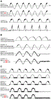
Extreme Delta Brush (EDB). A. This is a subset of +F, with abundant or continuous RDA+F or PDs+F (only if the PDs are blunt delta waves), where the fast activity has a stereotyped relationship to each delta wave. B. Extreme delta brush (EDB): RDA subtype. Examples of RDA that meet criteria for definite and possible EDB (A-C); with an example that does not (D), as the fast activity is part of the background (therefore this pattern does not count as RDA+F). C. Extreme Delta Brush (EDB): PD subtype. Examples of PDs (where the PDs are blunt delta waves) that meet criteria for definite and possible EDB. Example B is not EDB as the +F is not on the delta wave (i.e., it is not delta brushes). Example E is not +F as the fast activity is part of the background, therefore it can not be EDB.
FIG. 36.
FIG. 36.
A. Generalized Extreme Delta Brush (GEDB). GRDA+F also qualifies as definite GEDB if the RDA+F is abundant or continuous; and as possible GEDB if the RDA+F is occasional or frequent. B. Lateralized Extreme Delta Brush (LEDB). LRDA+F also qualifies as definite LEDB if the LRDA+F is abundant or continuous; and as possible LEDB if the LRDA+F is occasional or frequent. C. Bilateral Independent Extreme Delta Brush (BIEDB). BIRDA+F also qualifies as definite BIEDB if the BIRDA+F is abundant or continuous; and as possible EDB if the BIRDA+F is occasional or frequent.
FIG. 37.
FIG. 37.
Anterior-posterior (AP) lag.
FIG. 38.
FIG. 38.
Electrographic seizure (ESz).
FIG. 39.
FIG. 39.
Electroclinical seizure (ECSz).
FIG. 40.
FIG. 40.
Electroclinical seizure (ECSz)—for patients with previous known epileptic encephalopathy.
FIG. 41.
FIG. 41.
Brief Potentially Ictal Rhythmic Discharges (BIRDs). A. BIRDs with evolution, aka "evolving BIRDs" (a form of definite BIRDs). B. BIRDs with a similar morphology and location as interictal epileptiform discharges in the same patient (definite BIRDs). C. BIRDs with a similar morphology and location as seizures in the same patient (definite BIRDs). D. BIRDs that are sharply contoured but without the above features (possible BIRDs).
FIG. 42.
FIG. 42.
The Ictal-Interictal Continuum (IIC). Does not qualify as an electrographic seizure or electrographic status epilepticus but can be considered with any of the following features: A. Any PD or SW pattern that averages >1.0 Hz and ≤2.5 Hz over 10 seconds (>10 and ≤25 discharges in 10 seconds). B and Any PD or SW pattern that averages ≥0.5 Hz and ≤1.0 Hz over 10 seconds (≥ 5 and ≤10 discharges in 10 seconds), AND has a plus modifier or fluctuation. D and E. Any lateralized RDA (LRDA, BIRDA, UIRDA, MfRDA) averaging >1 Hz for ≥10 seconds (at least 10 waves in 10 seconds) with a plus modifier or fluctuation.

Comment in

References

    1. Hirsch LJ, Brenner RP, Drislane FW, et al. The ACNS subcommittee on research terminology for continuous EEG monitoring: proposed standardized terminology for rhythmic and periodic EEG patterns encountered in critically ill patients. J Clin Neurophysiol 2005;22:128–135. - PubMed
    1. Hirsch LJ, LaRoche SM, Gaspard N, et al. American clinical Neurophysiology Society’s Standardized Critical Care EEG Terminology: 2012 version. J Clin Neurophysiol 2013;30:1–27. - PubMed
    1. Gaspard N, Hirsch LJ, LaRoche SM, Hahn CD, Westover MB. Interrater agreement for critical care EEG terminology. Epilepsia 2014;55:1366–1373. - PMC - PubMed
    1. Lee JW, LaRoche S, Choi H, et al. Development and feasibility testing of a critical care EEG monitoring database for standardized clinical reporting and multicenter collaborative research. J Clin Neurophysiol 2016;33:133–140. - PMC - PubMed
    1. Gaspard N, Manganas L, Rampal N, Petroff OA, Hirsch LJ. Similarity of lateralized rhythmic delta activity to periodic lateralized epileptiform discharges in critically ill patients. JAMA Neurol 2013;70:1288–1295. - PubMed