. 2021 Jan-Dec;13(1):1-18.
doi: 10.1080/19490976.2020.1849996.

Salmonella effector SpvB aggravates dysregulation of systemic iron metabolism via modulating the hepcidin-ferroportin axis

Affiliations

Salmonella effector SpvB aggravates dysregulation of systemic iron metabolism via modulating the hepcidin-ferroportin axis

Qifeng Deng et al. Gut Microbes. 2021 Jan-Dec.

Abstract

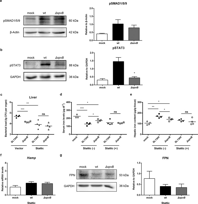
Iron withholding, an essential component of nutritional immunity, plays a fundamental role in host resistance to Salmonella infection. Our previous study showed that SpvB, an important pSLT-encoded cytotoxic effector, facilitated Salmonella pathogenesis within macrophages via perturbing cellular iron metabolism. However, the underlying mechanisms of SpvB in Salmonella-relevant disorders of systemic iron metabolism have not yet been identified. Here, we demonstrated that SpvB facilitated Salmonella to scavenge iron from the host by modulating the hepcidin-ferroportin axis, a key regulator of systemic iron metabolism. We observed that SpvB enhanced hepatic hepcidin synthesis in a STAT3-dependent manner, but not the BMP/SMAD pathway. This subsequently resulted in a reduction of the unique cellular iron exporter ferroportin, which facilitated hypoferremia and hepatic iron accumulation and ultimately countered the limitation of iron availability, thereby improving the chances of Salmonella survival and replication. Moreover, SpvB promoted the production of proinflammatory molecules associated with the infiltration of inflammatory cells via highly upregulating TREM-1 signaling. Our data supported a role of TREM-1 in SpvB-related dysregulation of host iron metabolism and suggested that targeting TREM-1 might provide a potential therapeutic strategy to prevent or alleviate Salmonella pathogenesis.

Keywords: Salmonella; SpvB; systemic iron metabolism; TREM-1; hepcidin–ferroportin axis.


Figures

Figure 1.
Figure 1.
SpvB effector protein is significant to S. typhimurium dysregulation of host iron metabolism. Streptomycin-pretreated mice were orally infected with 1 × 107 CFUs of either the WT or the ΔspvB mutant S. typhimurium strain. a) Hepatic bacterial load at 1 and 3 days post-infection was determined by plating. b) S. typhimurium-infected mice were administered either DFX dissolved in drinking water or the same volume of drinking water. Hepatic bacterial load at 3 days post-infection was determined by plating. c) Serum iron levels at 1 and 3 days post-infection were measured with a colorimetric assay. d) Hepatic iron content at 3 days post-infection was determined on the basis of a multiscan spectrum. e) Perls’ Prussian Blue staining of the liver at 3 days post-infection. Original magnification, ×40; scale bars: 50 μm. One of 3 representative histology experiments is shown. Statistical analysis was performed with IBM SPSS statistics 22. Data were compared with independent Student’s t-test. Values are expressed as the mean ± SEM, and statistically significant differences are indicated. *P< .05; **P< .01; ns, not significant
Figure 2.
Figure 2.
SpvB contributes to Salmonella-induced disorders of systemic iron metabolism by controlling the hepcidin-FPN axis. a-c) Streptomycin-pretreated mice were orally infected with 1 × 107 CFUs of either the WT or the ΔspvB mutant S. typhimurium strain. Hepatic Hamp levels at 1 day (a) or 3 days (b) post-infection were determined by quantitative PCR (n = 3–4 mice, respectively). c) Western blot analysis of whole liver lysates at 3 days post-infection with specific antibodies to FPN and the control GAPDH (n = 4 mice, respectively). Densitometric analysis of FPN relative to GAPDH protein and one of 4 representative western blot experiments are shown. d) Streptomycin-pretreated WT, Hamp+/− and Hamp−/− mice were orally infected with 1 × 107 CFUs of either the WT or the ΔspvB mutant S. typhimurium strain. Hepatic bacterial load at 3 days post-infection was determined by plating. Statistical analysis was performed with IBM SPSS statistics 22. Data were compared with independent Student’s t-test. Values are expressed as the mean ± SEM, and statistically significant differences are indicated. *P< .05; **P< .01; ns, not significant
Figure 3.
Figure 3.
SpvB increases hepatic hepcidin expression through the STAT3-dependent pathway. a, b) Streptomycin-pretreated mice were orally infected with 1 × 107 CFUs of either the WT or the ΔspvB mutant S. typhimurium strain and analyzed at 3 days post-infection. a) Western blot analysis of whole liver lysates with specific antibodies to pSMAD1/5/9 and the control β-Actin (n = 3 mice, respectively). Densitometric analysis of pSMAD1/5/9 relative to β-Actin protein and one of 3 representative western blot experiments are shown. b) Western blot analysis of whole liver lysates with specific antibodies to pSTAT3 and the control GAPDH (n = 3 mice, respectively). Densitometric analysis of pSTAT3 relative to GAPDH protein and one of 3 representative western blot experiments are shown. c-g) S. typhimurium-infected mice were administered i.p with either Stattic or the same volume of vector and analyzed at 3 days post-infection. c) Hepatic bacterial load was determined by plating. d) Serum iron levels were measured with a colorimetric assay. e) Hepatic iron content was determined on the basis of a multiscan spectrum. f) Hepatic Hamp levels were determined by quantitative PCR (n = 4 mice, respectively). g) Western blot analysis of whole liver lysates with specific antibodies to FPN and the control GAPDH (n = 3 mice, respectively). Densitometric analysis of FPN relative to GAPDH protein and one of 3 representative western blot experiments are shown. Statistical analysis was performed with IBM SPSS statistics 22. Data were compared with independent Student’s t-test. Values are expressed as the mean ± SEM, and statistically significant differences are indicated. *P< .05; **P< .01; ***P< .001; ns, not significant
Figure 4.
Figure 4.
SpvB promotes hepatic proinflammatory cytokine and chemokine expression in S. typhimurium infection. Streptomycin-pretreated mice were orally infected with 1 × 107 CFUs of either the WT or the ΔspvB mutant S. typhimurium strain and analyzed at 3 days post-infection. a-c) Hepatic Il1β (a), Tnfα (b) and Il6 (c) levels were determined by quantitative PCR (n = 4 mice, respectively). d) Western blot analysis of whole liver lysates with specific antibodies to IL6 and the control β-Actin (n = 3 mice, respectively). Densitometric analysis of IL6 relative to β-Actin protein and one of 3 representative western blot experiments are shown. e-g) Hepatic Ccl2 (e), Ccl3 (f) and Cxcl10 (g) levels were determined by quantitative PCR (n = 4 mice, respectively). Statistical analysis was performed with IBM SPSS statistics 22. Data were compared with independent Student’s t-test. Values are expressed as the mean ± SEM, and statistically significant differences are indicated. *P< .05; **P< .01; ***P< .001
Figure 5.
Figure 5.
SpvB increases inflammatory cell infiltration following S. typhimurium infection. Streptomycin-pretreated mice were orally infected with 1 × 107 CFUs of either the WT or the ΔspvB mutant S. typhimurium strain and analyzed at 3 days post-infection. a) Flow cytometric dot plots of hepatic non-parenchymal cells (n = 5 mice, respectively). b-d) Percentage of liver-infiltrated cell populations in S. typhimurium-infected mice. e) Histopathological analysis of the liver. Original magnification, ×20; scale bars: 100 μm. One of 3 representative histology experiments is shown. Statistical analysis was performed with IBM SPSS statistics 22. Data were compared with independent Student’s t-test. Values are expressed as the mean ± SEM, and statistically significant differences are indicated. *P< .05; ***P< .001
Figure 6.
Figure 6.
SpvB-mediated iron metabolic disorder is ameliorated by the TREM-1 inhibitor LP17. a, b) Streptomycin-pretreated mice were orally infected with 1 × 107 CFUs of either the WT or the ΔspvB mutant S. typhimurium strain and analyzed at 3 days post-infection. a) Hepatic Trem1 levels were determined by quantitative PCR (n = 4 mice, respectively). b) Western blot analysis of whole liver lysates with specific antibodies to TREM-1 and the control α-Tubulin (n = 3 mice, respectively). Densitometric analysis of TREM-1 relative to α-Tubulin protein and one of 3 representative western blot experiments are shown. c-i) S. typhimurium-infected mice were administered i.p with either LP17 or the same volume of vector and analyzed at 3 days post-infection. c) Hepatic bacterial load was determined by plating. d) Serum iron levels were measured with a colorimetric assay. e) Hepatic iron content was determined on the basis of a multiscan spectrum. f) Hepatic Hamp levels were determined by quantitative PCR (n = 4 mice, respectively). g) Western blot analysis of whole liver lysates with specific antibodies to FPN and the control GAPDH (n = 3 mice, respectively). Densitometric analysis of FPN relative to GAPDH protein and one of 3 representative western blot experiments are shown. h) Hepatic Il6 levels were determined by quantitative PCR (n = 4 mice, respectively). i) Western blot analysis of whole liver lysates with specific antibodies to pSTAT3 and the control GAPDH (n = 3 mice, respectively). Densitometric analysis of pSTAT3 relative to GAPDH protein and one of 3 representative western blot experiments are shown. Statistical analysis was performed with IBM SPSS statistics 22. Data were compared with independent Student’s t-test. Values are expressed as the mean ± SEM, and statistically significant differences are indicated. *P< .05; **P< .01; ***P< .001; ns, not significant
Figure 7.
Figure 7.
SpvB interferes macrophage iron metabolism when interacted with hepatocytes. a) HepG2 cells were infected with the WT, ΔspvB or c-spvB S. typhimurium strain at an MOI of 5, 10 or 20 for 8 h. HAMP levels were determined by quantitative PCR. b, c) Co-culture cells were infected with the WT, ΔspvB or c-spvB S. typhimurium strain at an MOI of 10 for 8 h. b) HAMP levels in co-cultured HepG2 cells were determined by quantitative PCR. c) Western blot analysis of co-cultured THP-1 macrophages lysates with specific antibodies to FPN and the control GAPDH. Densitometric analysis of FPN relative to GAPDH protein and one of 3 representative western blot experiments are shown. d) HepG2 cells with or without silencing of HAMP were co-cultured with THP-1 macrophages and then infected with the WT, ΔspvB or c-spvB S. typhimurium strain at an MOI of 10 for 8 h. Western blot analysis of co-cultured THP-1 macrophages lysates with specific antibodies to FPN and the control GAPDH. Densitometric analysis of FPN relative to GAPDH protein and one of 3 representative western blot experiments are shown. e, f) THP-1 cells with or without silencing of TREM-1 were co-cultured with HepG2 cells and then infected with the WT, ΔspvB or c-spvB S. typhimurium strain at an MOI of 10 for 8 h. e) HAMP levels in co-cultured HepG2 cells were determined by quantitative PCR. f) Western blot analysis of co-cultured THP-1 macrophages lysates with specific antibodies to FPN and the control GAPDH. Densitometric analysis of FPN relative to GAPDH protein and one of 3 representative western blot experiments are shown. Statistical analysis was performed with IBM SPSS statistics 22. The data were compared by ANOVA with Student-Newman-Keuls (S-N-K) correction. Values are expressed as the mean ± SEM of three independent experiments, and statistically significant differences are indicated. *P< .05; ns not significant
Figure 8.
Figure 8.
Model of SpvB in interfering with systemic iron metabolism. SpvB, an important pSLT-encoded cytotoxic effector, contributes to extensive and severe inflammation during S. typhimurium infection through activation of TREM-1 in macrophages. This subsequently induces hepatocyte hepcidin transcription via a IL6/JAK/STAT3-dependent manner. The induction of hepcidin expression in turn inhibits the sole iron exporter FPN, then results in an increased intramacrophage iron concentration and further facilitates hypoferremia and hepatic iron accumulation, which ultimately benefits for Salmonella survival and replication. MΦ, macrophage

Similar articles

Cited by

References

    1. Besser JM. Salmonella epidemiology: A whirlwind of change. Food Microbiol. 2018;71:55–18. doi:10.1016/j.fm.2017.08.018. - DOI - PubMed
    1. Fabrega A, Vila J. Salmonella enterica serovar Typhimurium skills to succeed in the host: virulence and regulation. Clin Microbiol Rev. 2013;26:308–341. - PMC - PubMed
    1. Guiney DG, Fierer J. The role of the spv genes in Salmonella pathogenesis. Front Microbiol. 2011;2:129. doi:10.3389/fmicb.2011.00129. - DOI - PMC - PubMed
    1. Kurita A, Gotoh H, Eguchi M, Okada N, Matsuura S, Matsui H, Danbara H, Kikuchi Y. Intracellular expression of the Salmonella plasmid virulence protein, SpvB, causes apoptotic cell death in eukaryotic cells. Microb Pathog. 2003;35(1):43–48. doi:10.1016/S0882-4010(03)00066-4. - DOI - PubMed
    1. Browne SH, Hasegawa P, Okamoto S, Fierer J, Guiney DG. Identification of Salmonella SPI-2 secretion system components required for SpvB-mediated cytotoxicity in macrophages and virulence in mice. FEMS Immunol Med Microbiol. 2008;52(2):194–201. doi:10.1111/j.1574-695X.2007.00364.x. - DOI - PubMed

Publication types

MeSH terms

LinkOut - more resources