. 2021 Jan 21;32(1):14.
doi: 10.1007/s10856-020-06483-6.

The effect of synthetic bone graft substitutes on bone formation in rabbit calvarial defects

Affiliations

The effect of synthetic bone graft substitutes on bone formation in rabbit calvarial defects

Nikola Saulacic et al. J Mater Sci Mater Med. .

Abstract

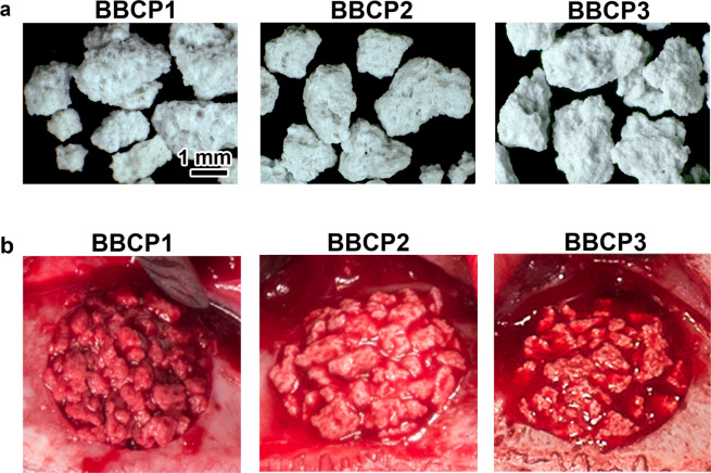
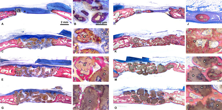
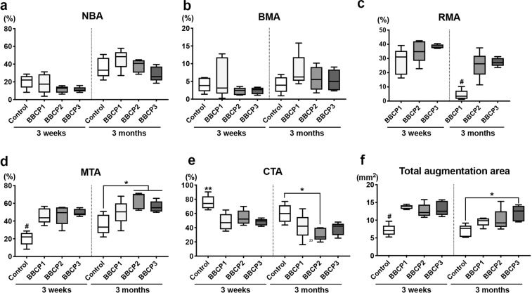
The aim of this study was to evaluate the influence of the intensity of the biomimetic hydroxyapatite (HA) coating of α-tricalcium phosphate (α-TCP) on biomaterial degradation and bone formation. Twenty-four female NZW rabbits of approximately 12 weeks of age were used. Critical size defects were randomly treated with 3%:97% HA:α-TCP (BBCP1), 12%:88% HA:α-TCP (BBCP2), and 23%:77% HA:α-TCP (BBCP3), respectively or sham. All defects were covered with a resorbable collagen membrane. Animals were euthanized after 3 and 12 weeks of healing and samples were investigated by micro-CT and histologic analysis. Ingrowth of newly formed woven bone from the original bone at 3-week healing period was observed in all samples. At the 12-week healing period, the new bone in the peripheral area was mainly lamellar and in the central region composed of both woven and lamellar bone. New bony tissue was found on the surface of all three types of granules and at the interior of the BBCP1 granules. Samples with 3% HA showed significantly less residual biomaterial in comparison to the other two groups. Furthermore, BBCP1 significantly promoted new bone area as compared to other three groups and more bone volume as compared to the control. Within its limitations, this study indicated the highest degradation rate in case of BBCP1 concomitant with the highest rate of bone formation. Hence, formation of new bone can be affected by the level of biomimetic HA coating of α-TCP.


Conflict of interest statement

The authors have no financial interest to declare in relation to the content of this article. Mr Claudio Zihlmann is employed by Geistlich Pharma AG (Wolhusen, Switzerland).

Figures

Fig. 1
Fig. 1
a Surface appearance of the particles of BBCP1, BBCP2, and BBCP3. The rough surfaces with pores were observed on each particle. b The images of the bone defects in rabbit parietal bone during surgeries. The materials were implanted in each defect, followed by mixing with autologous blood collected from auricular artery
Fig. 2
Fig. 2
ROIs for histomorphometry. ROI 1 (whole defect area), ROI 2 (middle area in a defect), and ROI 3 (lateral area in a defect) were investigated for each parameter
Fig. 3
Fig. 3
3D reconstructed micro-CT images of control (empty), BBCP1, BBCP2, and BBCP3 samples at 3 weeks and 3 months post surgery
Fig. 4
Fig. 4
a Bone volume (BV, mm3), b bone density (BD, relative % to initial bone at the defect site), c residual material volume (RMV, mm3), and d mineralized tissue volume (MTV, mm3, BV + RMV) at 3 weeks and 3 months post surgery. As a reference, control bone samples were used to show initial bone volume in the defect and initial bone density. *denotes significant difference between the groups (p < 0.05). #denotes significantly lower than any other modalities (p< 0.05)
Fig. 5
Fig. 5
Low (above) and high (under) magnified sectional images of the BBCP1 (a, d), BBCP2 (b, e), and BBCP3 (c, f) after fixing with blood. Blood cells including red blood cells and leucocytes infiltrated clearly only into the pores of the BBCP1
Fig. 6
Fig. 6
Representative histological images of control (a, b, i, j), BBCP1 (c, d, k, l), BBCP2 (e, f, m, n), and BBCP3 (g, h, o, p) groups at 3 weeks (left) and 3 months (right) of healing (toluidine blue and fuchsin staining). At 3 weeks, new bone is observed at the peripheral areas of the defect. The remnants of membranes are detectable covering the defect and the old bone. The outlined areas in the periphery of the defects (a, c, e, g) are magnified (b, d, f, h). All groups of granules (*) are showing contact to the newly formed woven bone (NB). At the 3 months of healing, new bone is seen in the central areas of the defect. The outlined areas (i, k, m, o) are magnified (j, l, n, p). Granules (*) are integrated in a newly formed lamellar bone (NB)
Fig. 7
Fig. 7
High magnification of the biomaterial at the center of defect after 3 weeks of healing. a At the interior of BBCP1 granules, multinucleated cells (*) and osteoblasts are identified inside the pores. b Direct contact of bone with BBCP2 granules is seen, with rare infiltration of the cells. c Small pores of the BBCP3 granules without cell infiltration
Fig. 8
Fig. 8
Center of the defect treated with BBCP1 (a), BBCP2 (b), and BBCP3 (c) granules after 3 weeks of healing. Granules are almost completely surrounded by a layer of multinuclear cells (arrowheads). Cell poor and highly vascularized loose connective tissue is observed between the granules
Fig. 9
Fig. 9
Periphery of defect treated with BBCP1 (a), BBCP2 (b), and BBCP3 (c) granules after 3 months of healing. Residual granules are embedded in the newly formed bone. Osteons (arrowheads) are observed within the newly formed lamellar bone and perforating the granules. The evidence for haematopoiesis with different degrees of cellularity is seen in all samples
Fig. 10
Fig. 10
Center of the defects treated with BBCP1 (a), BBCP2 (b), and BBCP3 (c) granules after 3 months of healing. Non-integrated granules are covered with a plenty of multinucleated giant cells (arrowheads) and a condensed fibrous tissue
Fig. 11
Fig. 11
The percentage of horizontal defect closure with bone at 3 weeks and 3 months post surgery in a whole defect (ROI 1), b middle area of the defect (ROI 2), and c lateral area of the defect (ROI 3). *denotes significant difference between the groups (p < 0.05). #denotes significantly lower than any other modalities (p < 0.05)
Fig. 12
Fig. 12
Histomorphometry for area parameter analysis at 3 weeks and 3 months post surgery in the whole defect (ROI 1); a new bone area (NBA, % to total augmentation area), b bone marrow area (BMA, % to total augmentation area), c residual material area (RMA, % to total augmentation area), d mineralized tissue area (MTA, NBA + RMA; % to total augmentation area), e connective tissue area (CTA, % to total augmentation area), and f total augmentation area (mm2). *denotes significant difference between the groups (p < 0.05). **denotes significantly higher than any other modalities (p < 0.05). #denotes significantly lower than any other modalities (p < 0.05)
Fig. 13
Fig. 13
Histomorphometry for area parameter analysis at 3 weeks and 3 months post surgery in the middle area of the defects (ROI 2); a new bone area (NBA, % to total augmentation area), b bone marrow area (BMA, % to total augmentation area), c residual material area (RMA, % to total augmentation area), d mineralized tissue area (MTA, NBA + RMA; % to total augmentation area), e connective tissue area (CTA, % to total augmentation area), and f total augmentation area (mm2). *denotes significant difference between the groups (p < 0.05). **denotes significantly higher than any other modalities (p < 0.05). #denotes significantly lower than any other modalities (p < 0.05)
Fig. 14
Fig. 14
Histomorphometry for area parameter analysis at 3 weeks and 3 months post surgery in the lateral area of the defects (ROI 3); a new bone area (NBA, % to total augmentation area), b bone marrow area (BMA, % to total augmentation area), c residual material area (RMA, % to total augmentation area), d mineralized tissue area (MTA, NBA + RMA; % to total augmentation area), e connective tissue area (CTA, % to total augmentation area), and f total augmentation area (mm2). *denotes significant difference between the groups (p < 0.05). **denotes significantly higher than any other modalities (p < 0.05). #denotes significantly lower than any other modalities (p < 0.05)

Similar articles

Cited by

References

    1. Miron RJ, Hedbom E, Saulacic N, Zhang Y, Sculean A, Bosshardt DD, et al. Osteogenic potential of autogenous bone grafts harvested with four different surgical techniques. J Dent Res. 2011;90:1428–33. doi: 10.1177/0022034511422718. - DOI - PubMed
    1. Nkenke E, Stelzle F. Clinical outcomes of sinus floor augmentation for implant placement using autogenous bone or bone substitutes: a systematic review. Clin Oral Implants Res. 2009;20:124–33. doi: 10.1111/j.1600-0501.2009.01776.x. - DOI - PubMed
    1. Moy P, Palacci P. Minor bone augmentation procedures. In: Palacci P, Ericsson I, editors. Esthetic implant dentistry. Soft and hard tissue management. Quintessence Publishing Co: IL.
    1. Wang RE, Lang NP. Ridge preservation after tooth extraction. Clin Oral Implants Res. 2012;23:147–56. doi: 10.1111/j.1600-0501.2012.02560.x. - DOI - PubMed
    1. Nissan J, Marilena V, Gross O, Mardinger O, Chaushu G. Histomorphometric analysis following augmentation of the posterior mandible using cancellous bone-block allograft. J Biomed Mater Res A. 2011;97:509–13. doi: 10.1002/jbm.a.33096. - DOI - PubMed

MeSH terms

Substances