. 2021 Mar 1;131(5):e138740.
doi: 10.1172/JCI138740.

Radiotherapy-exposed CD8+ and CD4+ neoantigens enhance tumor control

Affiliations

Radiotherapy-exposed CD8+ and CD4+ neoantigens enhance tumor control

Claire Lhuillier et al. J Clin Invest. .

Abstract

Neoantigens generated by somatic nonsynonymous mutations are key targets of tumor-specific T cells, but only a small number of mutations predicted to be immunogenic are presented by MHC molecules on cancer cells. Vaccination studies in mice and patients have shown that the majority of neoepitopes that elicit T cell responses fail to induce significant antitumor activity, for incompletely understood reasons. We report that radiotherapy upregulates the expression of genes containing immunogenic mutations in a poorly immunogenic mouse model of triple-negative breast cancer. Vaccination with neoepitopes encoded by these genes elicited CD8+ and CD4+ T cells that, whereas ineffective in preventing tumor growth, improved the therapeutic efficacy of radiotherapy. Mechanistically, neoantigen-specific CD8+ T cells preferentially killed irradiated tumor cells. Neoantigen-specific CD4+ T cells were required for the therapeutic efficacy of vaccination and acted by producing Th1 cytokines, killing irradiated tumor cells, and promoting epitope spread. Such a cytotoxic activity relied on the ability of radiation to upregulate class II MHC molecules as well as the death receptors FAS/CD95 and DR5 on the surface of tumor cells. These results provide proof-of-principle evidence that radiotherapy works in concert with neoantigen vaccination to improve tumor control.

Keywords: Antigen; Breast cancer; Immunology; Oncology; Radiation therapy.


Conflict of interest statement

Conflict of interest: SCF has received advisory or speaker compensation from AstraZeneca, Sanofi, EMD Serono, and Elekta, and research support from Bristol Myers Squibb, Merck, Eisai, Regeneron, Varian, and Janssen. SCF’s daughter is an employee of Pfizer. SD has received compensation for consultant/advisory services from Lytix Biopharma, Mersana Therapeutics, EMD Serono, and Ono Pharmaceutical, and research support from Lytix Biopharma and Nanobiotix. LG has received consulting fees from OmniSEQ, AstraZeneca, Inzen, and the Luke Heller TECPR2 Foundation, and he is member of the Scientific Advisory Committee of Boehringer Ingelheim, The Longevity Labs, and OmniSEQ.

Figures

Figure 1
Figure 1. Prediction and in vitro validation of MHC-I neoepitopes upregulated by radiotherapy in 4T1 breast cancer cells.
(A) Tumor neoantigen identification pipeline. Whole-exome and RNA sequencing data were used to identify mutations in genes expressed by 4T1 cells in vitro and in vivo and significantly upregulated by radiotherapy (RT). Candidate MHC-I binders were predicted using NetMHC and tested in binding assays. (B) RNA expression for Adgrf5, Dhx58, Cand1, and Raet1e genes as determined by RNA sequencing of 4T1 cells irradiated (3 × 8 Gy) or untreated (0 Gy) in vitro (n = 4 independent experiments). Data are expressed as mean ± SEM of normalized counts (log transformed). Comparisons between untreated and irradiated samples were made with unpaired 2-tailed Welch’s t test; *P < 0.05, ***P < 0.001. (C) MHC-I binding assay. RMA-S cells expressing either H2-Kd (left) or H2-Ld (right) were incubated with candidate peptides (50 or 100 μM) for 2 hours and then tested for MHC-I expression by flow cytometry. The binding capability of each peptide was calculated by normalization of the mean fluorescence intensity (MFI) of H2-Kd or H2-Ld in the presence of peptides compared with the MFI in the absence of peptides. HA515 peptide was used as positive control for H2-Kd, whereas AH1-A5 was used as positive control for H2-Ld. (D) Stability assay over time. RMA-S cells were loaded with peptides at 26°C for 1 hour and then transferred to 37°C before determination of H2-Kd (left) or H2-Ld (right) MFI by flow cytometry at the indicated times.
Figure 2
Figure 2. Identification of immunogenic neoepitopes in 4T1 cells.
(A) Experimental procedure. Mice were vaccinated twice with adjuvant alone, or with a combination of 2 neoepitopes binding to different alleles of MHC-I or MHC-II to avoid competition (75 μg each), as indicated (n = 4 per group). One week after the second vaccination, spleen and vaccine-draining lymph nodes were harvested and single-cell suspensions prepared for ex vivo stimulation experiments. (BE) Flow cytometry analysis of IFN-γ+TNF-α+ cells among CD8+ (B and C) or CD4+ (D and E) T cells from mice vaccinated as indicated in A and stimulated ex vivo with each peptide individually or in combination. Representative flow cytometry plots of IFN-γ/TNF-α intracellular staining in CD8+ (C) or CD4+ (E) T cells are shown after gating on viable CD3+ T cells. *P < 0.05, with Kruskal-Wallis and Dunn’s multiple-comparison tests. (F) Dose-response MHC-I binding assay comparing mutated and nonmutated (WT) DHX58 and CAND1 epitopes. RMA-S-Kd (left) or RMA-S-Ld (right) cells were incubated with increasing concentrations of the peptides for 2 hours before determination of H2-Kd or H2-Ld MFI by flow cytometry and normalization as in Figure 1C.
Figure 3
Figure 3. CAND1-specific CD8+ T cells are cytotoxic and kill preferentially irradiated 4T1 cells.
Mice were vaccinated twice with adjuvant alone (Control), with HA515, or with the 3 immunogenic neoepitopes (DHX58, CAND1, and ADGRF5-II; Neo-vax). (AC) One week after the last vaccination, mice were injected i.v. with CFSE-labeled target cells: CFSEhi cells were pulsed with indicated peptides, whereas CFSElo cells were unpulsed. Lymph nodes and spleen were harvested 24 hours later, and pulsed and unpulsed target cells were quantified by flow cytometry after gating on viable CD19+ CFSE+ cells. (A) Percentage of cytotoxicity toward HA515-paulsed target cells (n = 3 mice per group), calculated as indicated in Methods. *P < 0.05, with unpaired 2-tailed Welch’s t test. (B) Representative flow cytometry plots showing the killing of target cells in lymph nodes. (C) Percentage of cytotoxicity toward neoepitope-pulsed target cells determined in the lymph nodes and spleen (n = 3–7 mice per group). *P < 0.05, **P < 0.01, with Kruskal-Wallis and Dunn’s multiple-comparison tests. Data are pooled from 2 independent experiments. (D) Vaccine-draining lymph node cells were co-cultured with CFSEhi target cells pulsed with indicated neoepitopes, whereas CFSElo cells were pulsed with WT peptides. For HA515 peptide, CFSElo cells were unpulsed. MHC-I–blocking antibody was added as indicated. After incubation for 16–18 hours, cells were harvested for flow cytometry quantification of viable CD19+CFSE+ target cells, and the percentage of cytotoxicity toward mutated versus WT peptide-loaded targets was calculated. *P < 0.05, with unpaired 2-tailed Welch’s t test. Data are representative of 2 independent experiments. (EG) In vitro killing of 4T1 target cells was tested as described in the schema (E). (F) Representative flow plots after gating on viable CFSE+ cells. (G) Percentage of cytotoxicity toward irradiated cells versus untreated cells, calculated as described in Methods. **P < 0.01, with unpaired 2-tailed Welch’s t test. All data are expressed as mean ± SEM.
Figure 4
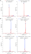
Figure 4. Targeted detection and quantification of neoepitopes and control epitope AH1 at the surface of tumor cells after irradiation.
SRM chromatograms of endogenous peptides (in red) and isotope-labeled peptides (in blue) for the AH1 epitope (SPSYVYHQF) (A), the CAND1 neoepitope (AYLSLLTQT) (B), and the ADGRF5-II neoepitope (STPMFSMSSPISRRF) (C). Detection and quantification of the peptides from untreated (left panels) or irradiated (right panels) 4T1 cells were performed by SRM analysis as described in Methods. Additionally, the MS/MS spectra for CAND1 and ADGRF5-II are shown in Supplemental Figures 3 and 4.
Figure 5
Figure 5. Vaccination with radiation-upregulated neoantigens enhances the antitumor immune response in combination with radiotherapy.
(A) Experimental schema. Mice were vaccinated twice with adjuvant alone (Control) or with the combination of 3 immunogenic neoepitopes (DHX58, CAND1, and ADGRF5-II; Neo-vax). One week after the second vaccination, 4T1 cells were injected s.c. in the right flank of mice. Vaccination was continued once a week. Tumors were irradiated with a dose of 8 Gy each on 3 consecutive days (RT). (B) Tumor volume measured over time after 4T1 cell inoculation (n = 9 per group). Statistical significance of Neo-vax + RT was assessed by repeated-measures 2-way ANOVA. (C) Tumor weight at day 25 (n = 8). (D) Number of clonogenic 4T1 lung metastases at day 25. Statistical significance was assessed by Kruskal-Wallis and Dunn’s multiple-comparison tests (n = 9). (EI) Flow cytometry analysis of the tumors at day 25 (n = 8). (E) Percentages of CD8+ T cells among viable CD45+ cells. (F) Representative flow plots of regulatory T cells (Tregs; CD25+FOXP3+) after gating on CD4+ T cells. (G) Absolute counts (normalized to tumor weight) of intratumoral Tregs and ratio of CD8+ cells to Tregs (H) or T-bet+ cells to Tregs (I). All data presented here are expressed as mean ± SEM. Comparisons between groups were made with Welch’s ANOVA and Dunnett’s T3 multiple-comparison tests, except in I, where the effect of Neo-vax was assessed by ordinary 2-way ANOVA. In all panels, *P < 0.05, **P < 0.01, ***P < 0.001, ****P < 0.0001.
Figure 6
Figure 6. Radiotherapy enhances T cell responses against radiation-upregulated neoantigens.
Mice were treated as in Figure 5A, and tumors and draining lymph nodes were harvested on day 25 for analysis. (AC) Representative flow plots (A), percentages (B), and absolute counts (C) of CAND1-dextramer–positive cells among intratumoral CD8+ T cells. Data are presented as mean ± SEM. Comparisons between groups were made with Kruskal-Wallis and Dunn’s multiple-comparison tests (n = 8); *P < 0.05, ***P < 0.001, ****P < 0.0001. (DF) Secreted IFN-γ in the supernatant of tumor-draining lymph node cells 48 hours after in vitro stimulation with DHX58 (D), CAND1 (E), and ADGRF5-II (F). Comparisons between all groups were made with Kruskal-Wallis and Dunn’s multiple-comparison tests; *P < 0.05, **P < 0.01, ***P < 0.001 (n = 6–8).
Figure 7
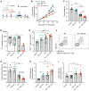
Figure 7. Therapeutic efficacy of the neoantigen vaccine in combination with radiotherapy.
(A) Experimental schema. Vaccination was started on day 4 after tumor inoculation and continued every 5–6 days thereafter. Tumors were irradiated with a dose of 8 Gy on 3 consecutive days (RT). (B and C) Tumor volume over time after 4T1 cell inoculation (n = 9 per group), shown as mean ± SEM (B) and as individual mouse curves (C). Statistical significance of RT + Neo-vax was assessed by repeated-measures 2-way ANOVA; **P < 0.01. (D) Mouse survival. ***P < 0.001, with log-rank test.
Figure 8
Figure 8. Both CD4+ and CD8+ neoantigen-specific T cells are required for the antitumor activity of the vaccine.
(A) Mice were vaccinated weekly with adjuvant alone (RT), all 3 neoepitopes (Neo-vax), or only 2 neoepitopes (CAND1/DHX58) starting 2 weeks before 4T1 cell inoculation. Tumors were irradiated with a dose of 8 Gy on days 12, 13, and 14. Tumor volume over time; *P < 0.05, with repeated-measures 2-way ANOVA (n = 8–9 mice per group). (B) Secreted IFN-γ was measured in the supernatant of tumor-draining lymph node cells harvested on day 31 and stimulated 48 hours with indicated peptides (CAND1, DHX58, ADGRF5-II, and AH1-A5). **P < 0.01, ***P < 0.001, with Kruskal-Wallis and Dunn’s multiple-comparison tests (n = 8). (C) In vitro killing of untreated or irradiated 4T1 target cells (as described in Figure 3E) using as effectors splenocytes harvested on day 31 and restimulated for 6 days with CAND1 or ADGRF5-II, followed by CD8+ or CD4+ T cell isolation, respectively. Ratios of untreated to irradiated cells were calculated as described in Methods. *P < 0.05, ***P < 0.001, with unpaired 2-tailed Student’s t test (n = 7). (D) Tumor volume over time of mice (n = 7–8 per group) treated as in A with adjuvant alone (RT) or with Neo-vax and RT. Some mice received CD4- or CD8-depleting antibodies starting 2 days before RT and maintained weekly; *P < 0.05, **P < 0.01, with repeated-measures 2-way ANOVA. All data presented are expressed as mean ± SEM.
Figure 9
Figure 9. CD4+ and CD8+ neoantigen-specific T cells kill tumor cells in an MHC- and death receptor–dependent manner.
(AC) Mice were vaccinated with adjuvant alone or with CAND1/ADGRF5-II weekly, starting 2 weeks before 4T1 cell inoculation. Tumors were irradiated with a dose of 8 Gy on days 12, 13, and 14. T cells were isolated from tumor-draining lymph nodes on day 20 and tested in an in vitro killing assay against untreated or irradiated 4T1 target cells (as described in Figure 3E), in the presence of blocking antibodies against MHC-I, MHC-II, FASL, and TRAIL. (A) Representative flow cytometry plots showing the 2 populations of 4T1 cells. (B and C) Ratios of untreated to irradiated cells calculated using absolute cell counts, as described in Methods. **P < 0.01, ***P < 0.001, with paired 2-tailed Student’s t test (n = 6–7). (D) Fas and Tnfrsf10b gene expression determined by RNA sequencing of 4T1 cells irradiated (3 × 8 Gy) or not (0 Gy) in vitro (n = 4 independent experiments). **P < 0.01, ***P < 0.001, with unpaired 2-tailed Welch’s t test. All data presented are expressed as mean ± SEM.

References

    1. Schumacher TN, Schreiber RD. Neoantigens in cancer immunotherapy. Science. 2015;348(6230):69–74. doi: 10.1126/science.aaa4971. - DOI - PubMed
    1. Rizvi NA, et al. Cancer immunology. Mutational landscape determines sensitivity to PD-1 blockade in non-small cell lung cancer. Science. 2015;348(6230):124–128. doi: 10.1126/science.aaa1348. - DOI - PMC - PubMed
    1. Le DT, et al. Mismatch repair deficiency predicts response of solid tumors to PD-1 blockade. Science. 2017;357(6349):409–413. doi: 10.1126/science.aan6733. - DOI - PMC - PubMed
    1. Snyder A, et al. Genetic basis for clinical response to CTLA-4 blockade in melanoma. N Engl J Med. 2014;371(23):2189–2199. doi: 10.1056/NEJMoa1406498. - DOI - PMC - PubMed
    1. Ott PA, et al. An immunogenic personal neoantigen vaccine for patients with melanoma. Nature. 2017;547(7662):217–221. doi: 10.1038/nature22991. - DOI - PMC - PubMed

Publication types

MeSH terms

Substances