. 2021 Jan 21;17(1):e1009233.
doi: 10.1371/journal.ppat.1009233. eCollection 2021 Jan.

SARS-CoV-2 variants with mutations at the S1/S2 cleavage site are generated in vitro during propagation in TMPRSS2-deficient cells

Affiliations

SARS-CoV-2 variants with mutations at the S1/S2 cleavage site are generated in vitro during propagation in TMPRSS2-deficient cells

Michihito Sasaki et al. PLoS Pathog. .

Abstract

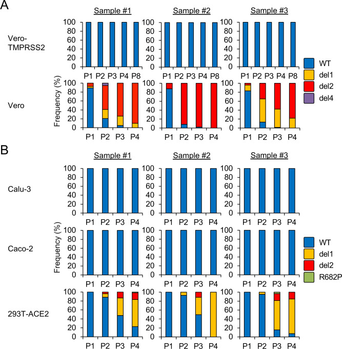
The spike (S) protein of Severe Acute Respiratory Syndrome-Coronavirus-2 (SARS-CoV-2) binds to a host cell receptor which facilitates viral entry. A polybasic motif detected at the cleavage site of the S protein has been shown to broaden the cell tropism and transmissibility of the virus. Here we examine the properties of SARS-CoV-2 variants with mutations at the S protein cleavage site that undergo inefficient proteolytic cleavage. Virus variants with S gene mutations generated smaller plaques and exhibited a more limited range of cell tropism compared to the wild-type strain. These alterations were shown to result from their inability to utilize the entry pathway involving direct fusion mediated by the host type II transmembrane serine protease, TMPRSS2. Notably, viruses with S gene mutations emerged rapidly and became the dominant SARS-CoV-2 variants in TMPRSS2-deficient cells including Vero cells. Our study demonstrated that the S protein polybasic cleavage motif is a critical factor underlying SARS-CoV-2 entry and cell tropism. As such, researchers should be alert to the possibility of de novo S gene mutations emerging in tissue-culture propagated virus strains.


Conflict of interest statement

I have read the journal's policy and the authors of this manuscript have the following competing interests: K.U., A.S., S.T., and T.S. are employees of Shionogi & Co., Ltd. Other authors have declared that no competing interests exist.

Figures

Fig 1
Fig 1. Isolation of SARS-CoV-2 S gene mutants.
(A) Schematic representation of SARS-CoV-2 S protein. Full-length S protein is cleaved into S1 and S2 proteins at the S1/S2 cleavage site. Functional domains (RBD, receptor binding domain; RBM, receptor binding motif) are highlighted. (B) Multiple amino acid sequence alignments focused on the S1/S2 cleavage site of wild type (WT) and isolated mutant viruses (del1, del2, del3 and R685H). Amino acid substitutions and deletions are shown as gray boxes, and the polybasic cleavage motif (RARR) at the S1/S2 cleavage site is highlighted in red. A red arrowhead indicates S1/S2 cleavage site. (C) Plaque formation for SARS-CoV-2 WT and isolated mutants grown in Vero-TMPRSS2 cells on 6 well plates. (D) Detection of virus S and N proteins in Vero-TMPRSS2 cells infected with WT or isolated mutants. The full-length S and cleaved S2 proteins are indicated by closed and open arrowheads, respectively.
Fig 2
Fig 2. Infection and growth of SARS-CoV-2 S gene mutants in different cell lines.
(A) Vero, Vero-TMPRSS2, Caco-2 and Calu-3 cells were inoculated with SARS-CoV-2 WT or S gene mutants at a multiplicity of infection (MOI) of 1. At 24 h post infection, cells were stained with anti-SARS-CoV-2 S antibody (green) and Hoechst 33342 nuclear dye (blue); scale bars, 50 μm. White arrowheads indicate cell syncytia. (B) Cells were inoculated with SARS-CoV-2 WT or S gene mutants at an MOI of 0.1. Culture supernatants were harvested at 24, 48 and 72 h after inoculation. Virus titration was performed by plaque assay. The values shown are mean ± standard deviation (SD) of triplicate samples. One-way analysis of variance with Dunnett's test was used to determine the statistical significance of the differences in virus titer between the WT and S gene mutants at 72 h after inoculation. *p < 0.05, **p < 0.01.
Fig 3
Fig 3. Infection and growth of SARS-CoV-2 S gene mutants in 293T and 293T-ACE2 cells.
(A) 293T and 293T-ACE2 cells were inoculated with SARS-CoV-2 WT or S gene mutants at an MOI of 1. At 24 h after inoculation, cells were stained with anti-SARS-CoV-2 S antibody (green) and Hoechst 33342 nuclear dye (blue); scale bars, 50 μm. (B) Cells were infected with SARS-CoV-2 WT or S gene mutants at an MOI of 0.1. Culture supernatants were harvested at 24, 48 and 72 h after inoculation and titration of infectious virus was determined by plaque assay. The values shown are means ± SD of triplicate samples. One-way analysis of variance with Dunnett's test was used to determine the statistical significance of the differences in virus titer between the WT and S gene mutants at 72 h after inoculation. **p < 0.01.
Fig 4
Fig 4. Impact of biochemical inhibitors on cellular entry of SARS-CoV-2 S gene mutants.
(A) Vero-TMPRSS2 cells were infected with SARS-CoV-2 WT or del2 mutant in the presence of varying concentrations of the TMPRSS2 inhibitor, camostat, or the cathepsin B/L inhibitor, E-64d, for 1h. At 6 h post-inoculation, the relative levels of viral N protein RNA were evaluated quantitatively by qRT-PCR. (B) Vero-TMPRSS2 and Vero cells were infected with SARS-CoV-2 WT or S gene mutants in the presence of 50 μM camostat and/or 25 μM E-64d for 1 h. At 6 h post-inoculation, the relative levels of viral N protein RNA were quantified by qRT-PCR. Cellular β-actin mRNA levels were used as reference controls. The values shown are mean ± SD of triplicate samples. One-way analysis of variance with Dunnett's test was used to determine the statistical significance between the responses to treatment with inhibitors and the no-treatment controls; *p < 0.05, **p < 0.01, ***p < 0.001.
Fig 5
Fig 5. Frequencies of S gene mutants detected during SARS-CoV-2 propagation.
SARS-CoV-2 was serially passaged in (A) Vero-TMPRSS2, Vero cells or (B) Calu-3, Caco-2 and 293T-ACE2 cells, each with three biological replicates. Nucleotide sequence diversity at viral S1/S2 cleavage site was determined by deep-sequencing.

References

    1. (WHO) WHO. Coronavirus disease (COVID-19) pandemic 2020 [cited 2020 9th November]. Available from: https://www.who.int/emergencies/diseases/novel-coronavirus-2019.
    1. Naqvi AAT, Fatima K, Mohammad T, Fatima U, Singh IK, Singh A, et al. Insights into SARS-CoV-2 genome, structure, evolution, pathogenesis and therapies: Structural genomics approach. Biochim Biophys Acta Mol Basis Dis. 2020;1866(10):165878 Epub 2020/06/13. 10.1016/j.bbadis.2020.165878 - DOI - PMC - PubMed
    1. Wang YT, Landeras-Bueno S, Hsieh LE, Terada Y, Kim K, Ley K, et al. Spiking Pandemic Potential: Structural and Immunological Aspects of SARS-CoV-2. Trends Microbiol. 2020. Epub 2020/05/20. 10.1016/j.tim.2020.05.012 - DOI - PMC - PubMed
    1. Walls AC, Park YJ, Tortorici MA, Wall A, McGuire AT, Veesler D. Structure, Function, and Antigenicity of the SARS-CoV-2 Spike Glycoprotein. Cell. 2020;181(2):281–92.e6. Epub 2020/03/09. 10.1016/j.cell.2020.02.058 - DOI - PMC - PubMed
    1. Wrapp D, Wang N, Corbett KS, Goldsmith JA, Hsieh CL, Abiona O, et al. Cryo-EM structure of the 2019-nCoV spike in the prefusion conformation. Science. 2020;367(6483):1260–3. Epub 2020/02/19. 10.1126/science.abb2507 - DOI - PMC - PubMed

Publication types

Substances