. 2021 Feb 4;184(3):628-642.e10.
doi: 10.1016/j.cell.2020.12.027. Epub 2020 Dec 23.

Compromised SARS-CoV-2-specific placental antibody transfer

Affiliations

Compromised SARS-CoV-2-specific placental antibody transfer

Caroline Atyeo et al. Cell. .

Abstract

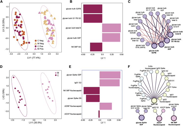
SARS-CoV-2 infection causes more severe disease in pregnant women compared to age-matched non-pregnant women. Whether maternal infection causes changes in the transfer of immunity to infants remains unclear. Maternal infections have previously been associated with compromised placental antibody transfer, but the mechanism underlying this compromised transfer is not established. Here, we used systems serology to characterize the Fc profile of influenza-, pertussis-, and SARS-CoV-2-specific antibodies transferred across the placenta. Influenza- and pertussis-specific antibodies were actively transferred. However, SARS-CoV-2-specific antibody transfer was significantly reduced compared to influenza- and pertussis-specific antibodies, and cord titers and functional activity were lower than in maternal plasma. This effect was only observed in third-trimester infection. SARS-CoV-2-specific transfer was linked to altered SARS-CoV-2-antibody glycosylation profiles and was partially rescued by infection-induced increases in IgG and increased FCGR3A placental expression. These results point to unexpected compensatory mechanisms to boost immunity in neonates, providing insights for maternal vaccine design.

Keywords: Fc-receptor; SARS-CoV-2; antibodies; fucose; glycosylation; hypergammablobulinemia; infection; inflammation; placental transfer; pregnancy; trimester.


Conflict of interest statement

Declaration of interests G.A. is the founder of Seromyx. K.J.G. has consulted for BillionToOne, Quest Diagnostics, Illumina, and Aetion. A.A.B. has consulted for Microchips Biotech and is also a Scientific Advisory Board Member for Reproductive Health Investors Alliance. D.P. owns stock in Gilead Sciences, BioNano Genomics, Biogen, Bluebird Bio, ImmunoGen, Pfizer, and Bristol-Myers Squibb. Any opinion, findings, and conclusions or recommendations expressed in this material are those of the authors(s) and do not necessarily reflect the views of the National Science Foundation.

Figures

None
Graphical abstract
Figure 1
Figure 1
Inefficient placental transfer of SARS-CoV-2 antibodies (A) The dot plots show the relative hemagglutinin- (HA) (left) and pertactin- (PTN) (right) specific IgG1 titers for both COVID-negative (COVID, orange) and COVID-positive (COVID+, pink) maternal-cord pairs. The dotted line indicates background PBS levels. Significance was determined by a Wilcoxon signed-rank test, p < 0.05, ∗∗∗p < 0.001, ∗∗∗∗p < 0.0001. (B) The dot plot shows the IgG1 titer against SARS-CoV-2 receptor binding domain (RBD, left), spike (S) (middle), and nucleocapsid (N) (right). The lines connect maternal:cord pairs. The dotted line shows the PBS background level. Significance was determined by Wilcoxon signed-rank test, p < 0.05. (C) The dot plots show the transfer ratio of HA, PTN, RBD, S, and N antibodies. The transfer ratio of COVID dyads are shown in orange, whereas the transfer ratios of COVID+ dyads are shown in pink. The horizontal dotted line indicates a 100% transfer efficiency (equivalent levels across both compartments). Significance was determined by using a one-way ANOVA followed by Tukey’s multiple comparisons test, where “ha” indicates a significant difference compared to the COVID+ HA transfer ratio and “p” indicates a significant difference compared to the COVID+ PTN transfer ratio. ∗∗p < 0.01, ∗∗∗p < 0.001, ∗∗∗∗p < 0.0001. (D) The heatmap shows the median PBS-background corrected transfer ratio of HA, PTN, RBD, S, and N across all antibody subclasses and FcR binding profiles. See also Figures S1, S2, and S3.
Figure S1
Figure S1
HA-specific antibodies are increased in the cord across subclasses and FcR-binding profiles, related to Figure 1 A-C. The dot plots show the relative HA-specific IgG2 (A, right), IgG3 (A, left), FcRn-binding (B), and FcgR-binding (C) in maternal plasma (M) and cord blood (C) for COVID- dyads (left, orange) and COVID+ dyads (right, pink). Lines connect maternal (M):cord (C) pairs. Significance was determined by a Wilcoxon signed rank test: ns is not significant, p < 0.05, ∗∗p < 0.01, ∗∗∗p < 0.001, ∗∗∗∗p < 0.0001.
Figure S2
Figure S2
SARS-CoV-2-targeting antibodies are transferred inefficiently to the cord across subclasses and FcR binding, related to Figure 1 A. The dot plots show the titer of IgG1 against SARS-CoV-1 S1 and S2 in SARS-CoV-2 infected dyads. Lines connect maternal (M):cord (C) pairs. Significance was determined by a Wilcoxon signed rank test, p < 0.05, ∗∗p < 0.01, ∗∗∗p < 0.001, ∗∗∗∗p < 0.0001. B. The heatmap shows the median transfer ratio of SARS-CoV-2 S1 and S2-targeting IgG and FcR-binding. C-H. The dot plots show the titer of IgG2 (C), IgG3 (D), and FcR binding (E-H) against RBD, spike (S), nucleocapsid (N), S1 and S2 in maternal plasma (M) and cord blood. Lines connect maternal (M):cord (C) pairs. Significance was determined by a Wilcoxon signed rank test, p < 0.05, ∗∗p < 0.01, ∗∗∗p < 0.001, ∗∗∗∗p < 0.0001.
Figure S3
Figure S3
The effect of time on SARS-CoV-2-specific antibody transfer, related to Figures 1 and 4 A-B. The dot plots show the relative IgG titer for RBD, S, N and HA in maternal plasma and cord blood in which the mother was infected during the third (A) or second (B) trimester. Lines connect mother:cord dyads. The data is represented as the absorbance at 450 nm subtracted by the reference absorbance read at 570 nm. The data was background corrected using plasma that was negative for SARS-CoV-2 antibodies. (C). Transfer ratios for IgG1 and FcRn were plotted according to time from last vaccination for HA (left) or from symptom onset or positive test for SARS-CoV-2 S and N (right), using a median split based on time. Logistic regression was performed to calculate the probability of each transfer ratio to be greater than 1 based on time (green line). D. An orthogonal partial least square regression (OPLSR) was built using SARS-CoV-2-specific antibody features and spike-specific glycan data with IgG1 spike transfer ratio as the outcome variable. The scores plot is colored based on IgG1 spike transfer ratio (left). When time (or days since symptom onset/positive test result) was added to the Elastic Net reduced feature set, the loadings of the previously selected features changed minimally and the Elastic Net selected features, rather than time elapsed since infection, continued to drive IgG1 spike transfer ratio prediction (right).
Figure 2
Figure 2
Functional HA-, but not SARS-CoV-2-, specific antibodies are transferred efficiently across the placenta (A–D) The dot plots illustrate antibody-dependent monocyte phagocytosis (ADCP, A), natural killer cell (NK) activation (B), antibody-dependent neutrophil phagocytosis (ADNP, C), and antibody-dependent complement deposition (ADCD, D) (PBS background corrected) in maternal plasma (M) and cord-blood plasma (C) against hemagglutinin (HA). Lines connect maternal:cord pairs. COVID dyads are represented in orange, and COVID+ dyads are represented in pink. Significance was determined by a Wilcoxon signed-rank test, p < 0.05, ∗∗p < 0.01, ∗∗∗p < 0.001, ∗∗∗∗p < 0.0001. (E–H) The dot plots show the ADCP (E), NK activation (F), ADNP (G), and ADCD (H) activity (PBS background corrected) in maternal blood (M) and cord plasma (C) for the SARS-CoV-2 antigens RBD, spike (S), and nucleocapsid (N). Lines connect maternal:cord dyads. Significance was determined by a Wilcoxon signed-rank test, p < 0.05, ∗∗p < 0.01. (I–L) The box-and whisker plots show the transfer-ratios for ADCP (I), NK activation (J), ADNP (K), and ADCD (L) against HA, RBD, S, and N. For HA, COVID dyads are represented in orange and COVID+ dyads are shown in pink. For each SARS-CoV-2 antigen, significance was determined against the HA activity using the matched COVID+ dyad. A one-way ANOVA followed by Tukey’s multiple comparison test was performed to determine significance, p < 0.05, ∗∗p < 0.01, ∗∗∗p < 0.001.
Figure 3
Figure 3
Spike-specific Fc-glycan profiles diverge from bulk antibody profiles (A) The figure represents a typical Fc glycan. The solid lines between sugars represent the linkages that are always present, whereas the dotted lines represent sugars that can be added. (B) An orthogonal partial least-squares discriminant analysis (OPLSDA) model was built using bulk glycan features from COVID (orange) and COVID+ (pink) maternal plasma samples. This orthogonal approach ensures that maximal variance between groups is captured in the first latent variable (LV1). The dot plot shows the distribution of each maternal sample, with ellipses encompassing the 95% confidence interval for each group. (C) The dot plot shows the percent of each glycoform in bulk antibodies in maternal (M) and cord (C) samples. The glycoforms are denoted as follows: agalactosylated (G0), monogalactosylated (G1), digalactosylated (G2), fucosylated (F), sialylated (S), and bisected n-acetyl-glucosamine (GlcNAc) (B). Connected lines signify maternal:cord dyads. Significance was determined using a Wilcoxon signed-rank test, p < 0.05, ∗∗p < 0.01, ∗∗∗p < 0.001, ∗∗∗∗p < 0.0001. (D) A multi-level principal component analysis (m-PCA) was built using bulk (pink) and spike-specific (purple) antibody glycan data from COVID+ mothers for which matched data were available. Each maternal sample is represented as a dot on the scores plot (left). The bar graph (right) shows the loadings on principal component 1 (PC1) of each glycan feature. (E) The dot plot shows the percentage of each glycoform in maternal bulk (pink) and spike-specific (purple) antibodies. The lines represent glycan data from the same mother. Significance was determined using a Wilcoxon signed-rank test, p < 0.05, ∗∗p < 0.01, ∗∗∗p < 0.001, ∗∗∗∗p < 0.0001. (F) The dot plot shows the percent of each glycoform in spike-specific antibodies in maternal (M) and cord (C) samples. Significance was determined using a Wilcoxon signed-rank test (G) The boxplots show the transfer ratio of each glycoform for bulk (orange/pink) and spike-specific (purple) antibodies. For bulk glycan data, COVID dyads are represented in orange and COVID+ dyads are represented in pink. Spike (S)-specific glycan data are shown in purple. Significance was determined by a Mann-Whitney test between COVID+ bulk and S-specific antibodies for each glycan feature, p < 0.05, ∗∗p < 0.01, ∗∗∗p < 0.001, ∗∗∗∗p < 0.0001. See also Figure S4.
Figure S4
Figure S4
Multivariate model validation, related to Figures 3 and 4 A-C. ROC curves corresponding to m-OPLSDA models in (A) Figure 3B, (B) Figures 4A–4C, and (C) Figures 4D–4F. 100 trials of 5-fold CV were run for each model, illustrated by colored curves. The ROC curve corresponding with the median AUC score was plotted in black with the numerical score reported in the legend. The blue dashed line indicates the classification threshold for a random process. D. Table of performance metrics for each OPLSDA model, including mean accuracy score (left) and comparison models generated with random features (center) or permuted disease state labels (right). In each case, 100 trials were performed with 5-fold cross validation. (E). Plot of actual-versus-predicted transfer ratios for the OPLSR models Figure S3D. The solid line represents the linear regression of the data and the shaded area represents the standard deviation (SD) of the slope of the regression line when leave-one-out cross validation was performed. Slope approaching the value of 1 indicates good model performance.
Figure 4
Figure 4
Placental sieving signatures of HA- and spike-specific antibodies (A) Multi-level orthogonal partial least square discriminant analysis (m-OPLSDA) was performed to distinguish between profiles from mother and cord plasma using HA, PTN, and bulk glycan data. Triangular and circular data points on the scores plot represent mother and cord samples, respectively. Infection status is indicated by color (orange representing COVID and pink representing COVID+ individuals). The ellipses show the 95% confidence interval for the mother/cord populations. (B) The bar graph illustrates the loadings on latent variable 1 (LV1) of the Elastic Net-selected antibody features from the m-OPLSDA shown in (A). The color indicates the group in which each feature is enriched (dark pink for cord samples, light pink for maternal plasma). (C) A pairwise spearman correlation test was performed to determine features that correlate with the Elastic Net-selected features (bolded nodes) selected for the m-OPLSDA model shown in (A). Features with correlation of coefficients >|0.75| and p < 0.05 are shown in the networks. Positive correlations are shown in purple, and negative correlations are shown in pink. Nodes are colored by features type, with glycan data shown in purple, antibody titer shown in yellow, and FcR binding shown in blue. (D) A m-OPLSDA model was built to classify COVID+ mother and cord plasma using SARS-CoV-2-specific antibody features and spike-specific glycan data. Only samples for which spike-specific glycan data were available were included in the model. The scores plot shows the separation between the cord samples (dark pink) and the maternal samples (light pink), where each dot represents a sample. The ellipses show the 95% confidence interval for each group. (E) The bar graph shows the loadings on latent variable 1 (LV1) of the Elastic Net-selected features for the m-OPLSDA model built in (D). The color indicates the group in which each feature is enriched (dark pink for cord samples, light pink for maternal samples). (F) A pairwise spearman correlation test was performed to determine features that were correlated with the Elastic Net-selected features (bold nodes) that were selected for the m-OPLSDA model shown in (D). Features with correlation of coefficients >|0.75| and p < 0.05 are shown in the networks. Positive correlations are shown in purple and negative correlations are shown in pink. Nodes are colored by feature type, with glycan data shown in purple, antibody titer shown in yellow, and FcR binding shown in blue. See also Figures S3 and S4.
Figure 5
Figure 5
Elevated IgG compensates for glycosylation changes to improve SARS-CoV-2-specific antibody transfer (A and B) A total IgG ELISA was run on COVID (orange) and COVID+ (pink) maternal plasma (A) and cord blood (B). Significance was determined by a Mann-Whitney test. p < 0.05, ∗∗p < 0.01, ∗∗∗p < 0.001, ∗∗∗∗p < 0.0001. (C) The boxplots show the transfer ratio, calculated as cord OD/maternal OD. The dotted line shows a transfer ratio of 1. Significance was determined by a Mann-Whitney test. (D–F) The dot plots illustrate the correlation between IgG titer and IgG1 HA (D and E) or IgG1 Spike (F) transfer ratio in COVID negative (orange) and COVID positive (pink) individuals. The direction of correlation is indicated by the color of the regression line (neg, blue; pos, purple). (G) 2-feature linear regression models were built to determine which feature, in combination with total IgG titer, was most predictive of SARS-CoV-2-specific antibody transfer ratio. The boxplots show the adjusted R2 values for the top performing models over 10 leave-one-out cross-validated trials. (H) The dots represent the adjusted R2 values of three of the top 2-feature models (G) to each of the individual features predictive performance of SARS-CoV-2-specific antibody transfer. For the 1-feature regression models, data points are colored by the direction of correlation with IgG1 spike transfer ratio (neg, blue; pos, purple). Error bars illustrate standard deviation of adjusted R2 after 10 rounds of leave-one-out cross-validation. (I) The dot plots represent the ratio of percent fucosylated to total percent for each galactose feature. For each galactose glycoform, the data were normalized by dividing by the average ratio of the bulk COVID. Significance was only calculated between bulk COVID+ and spike-specific antibodies. Significance was determined by Mann-Whitney test, p < 0.05, ∗∗p < 0.01, ∗∗∗p < 0.001, ∗∗∗∗p < 0.0001.
Figure 6
Figure 6
Increased FcRn/FCGR3A colocalization following SARS-CoV-2 infection (A) Placental tissue sections from COVID+ and COVID mothers were stained for FcRn (purple). FCGR3A (red), and placental alkaline phosphatase (PLAP, green), a trophoblast marker. (B) The dot plots represent the intensity of FcRn, FCGR3, and FcRn/FCGR3 colocalization in placental tissues. Significance was determined by an unpaired Student’s t test with Welch’s correction, ∗∗p < 0.01.

References

    1. Abu Raya B., Srugo I., Kessel A., Peterman M., Bader D., Gonen R., Bamberger E. The effect of timing of maternal tetanus, diphtheria, and acellular pertussis (Tdap) immunization during pregnancy on newborn pertussis antibody levels - a prospective study. Vaccine. 2014;32:5787–5793. - PubMed
    1. Ackerman M.E., Moldt B., Wyatt R.T., Dugast A.S., McAndrew E., Tsoukas S., Jost S., Berger C.T., Sciaranghella G., Liu Q. A robust, high-throughput assay to determine the phagocytic activity of clinical antibody samples. J. Immunol. Methods. 2011;366:8–19. - PMC - PubMed
    1. Ackerman M.E., Crispin M., Yu X., Baruah K., Boesch A.W., Harvey D.J., Dugast A.S., Heizen E.L., Ercan A., Choi I. Natural variation in Fc glycosylation of HIV-specific antibodies impacts antiviral activity. J. Clin. Invest. 2013;123:2183–2192. - PMC - PubMed
    1. Alter G., Ottenhoff T.H.M., Joosten S.A. Antibody glycosylation in inflammation, disease and vaccination. Semin. Immunol. 2018;39:102–110. - PMC - PubMed
    1. Alzamora M.C., Paredes T., Caceres D., Webb C.M., Valdez L.M., La Rosa M. Severe COVID-19 during Pregnancy and Possible Vertical Transmission. Am. J. Perinatol. 2020;37:861–865. - PMC - PubMed

Publication types