

Protective effect of Rosuvastatin on Azithromycin induced cardiotoxicity in a rat model
- PMID: 33476632
- PMCID: PMC7816566
- DOI: 10.1016/j.lfs.2021.119099
Protective effect of Rosuvastatin on Azithromycin induced cardiotoxicity in a rat model
Abstract
Aims: Azithromycin is widely used broad spectrum antibiotic recently used in treatment protocol of COVID-19 for its antiviral and immunomodulatory effects combined with Hydroxychloroquine or alone. Rat models showed that Azithromycin produces oxidative stress, inflammation, and apoptosis of myocardial tissue. Rosuvastatin, a synthetic statin, can attenuate myocardial ischemia with antioxidant and antiapoptotic effects. This study aims to evaluate the probable protective effect of Rosuvastatin against Azithromycin induced cardiotoxicity.
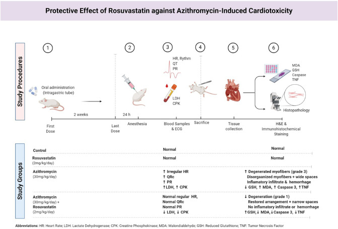
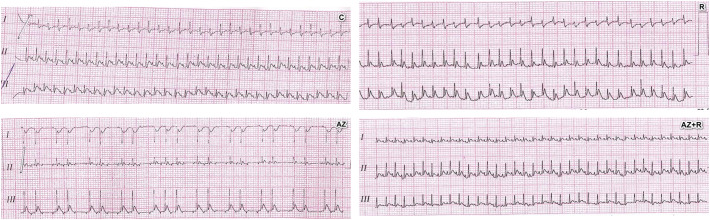
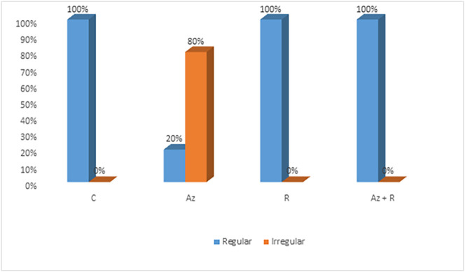
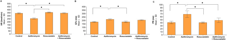
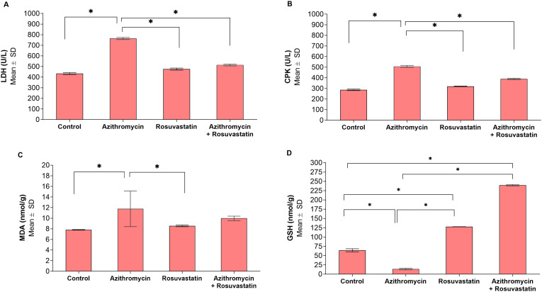
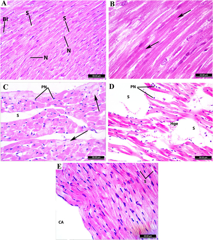
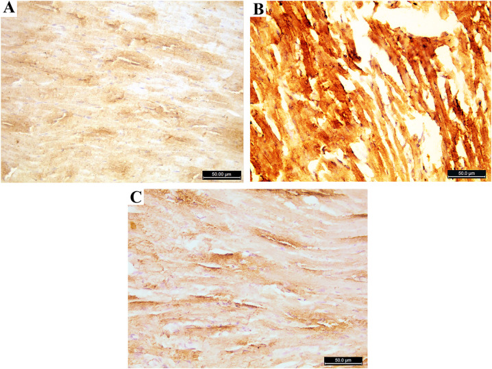
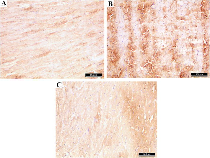
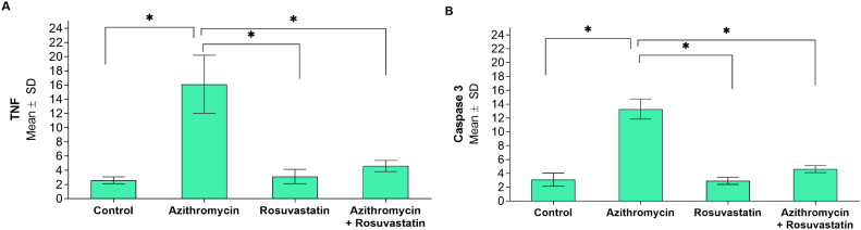
Main method: Twenty adult male albino rats were divided randomly into four groups, five rats each control, Azithromycin, Rosuvastatin, and Azithromycin +Rosuvastatin groups. Azithromycin 30 mg/kg/day and Rosuvastatin 2 mg/kg/day were administrated for two weeks by an intragastric tube. Twenty-four hours after the last dose, rats were anesthetized and the following measures were carried out; Electrocardiogram, Blood samples for Biochemical analysis of lactate dehydrogenase (LDH), and creatine phosphokinase (CPK). The animals sacrificed, hearts excised, apical part processed for H&E, immunohistochemical staining, and examined by light microscope. The remaining parts of the heart were collected for assessment of Malondialdehyde (MDA) and Reduced Glutathione (GSH).
Key findings: The results revealed that Rosuvastatin significantly ameliorates ECG changes, biochemical, and Oxidative stress markers alterations of Azithromycin. Histological evaluation from Azithromycin group showed marked areas of degeneration, myofibers disorganization, inflammatory infiltrate, and hemorrhage. Immunohistochemical evaluation showed significant increase in both Caspase 3 and Tumor necrosis factor (TNF) immune stain. Rosuvastatin treated group showed restoration of the cardiac muscle fibers in H&E and Immunohistochemical results.
Significance: We concluded that Rosuvastatin significantly ameliorates the toxic changes of Azithromycin on the heart.
Keywords: Azithromycin; Heart; Rat; Rosuvastatin.
Copyright © 2021 Elsevier Inc. All rights reserved.
Conflict of interest statement
There are no conflicts of interest.
Figures
References
-
- Li X., Wang M., Liu G., Ma J., Li C. Association of macrolides with overall mortality and cardiac death among patients with various infections: a meta-analysis. European Journal of Internal Medicine. 2016;28:32–37. - PubMed
-
- Du X., Zuo X., Meng F., Wu F., Zhao X., Li C., et al. Combinatorial screening of a panel of FDA-approved drugs identifies several candidates with anti-Ebola activities. Biochem. Biophys. Res. Commun. 2020;522:862–868. - PubMed
MeSH terms
Substances
LinkOut - more resources
Full Text Sources
Other Literature Sources
Medical
Research Materials
