. 2021 Jan-Jun:296:100306.
doi: 10.1016/j.jbc.2021.100306. Epub 2021 Jan 19.

SARS-CoV-2 infects cells after viral entry via clathrin-mediated endocytosis

Affiliations

SARS-CoV-2 infects cells after viral entry via clathrin-mediated endocytosis

Armin Bayati et al. J Biol Chem. 2021 Jan-Jun.

Abstract

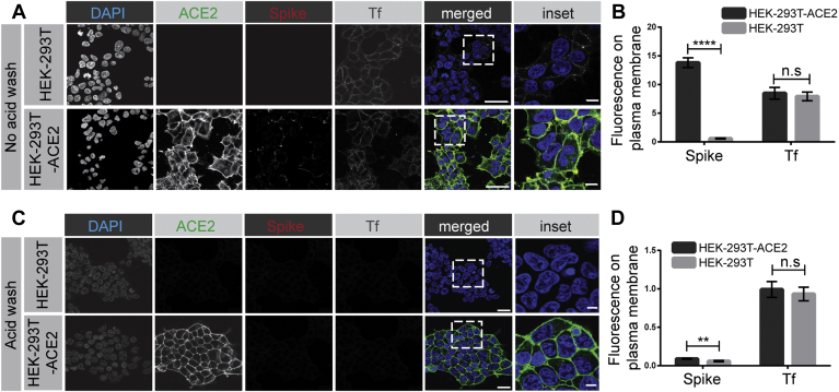
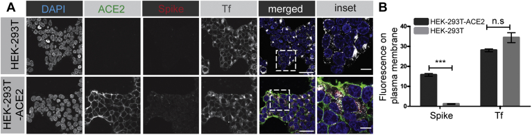
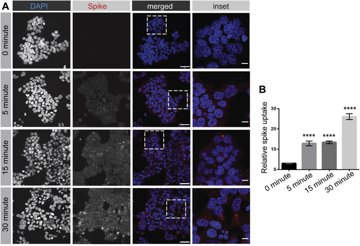
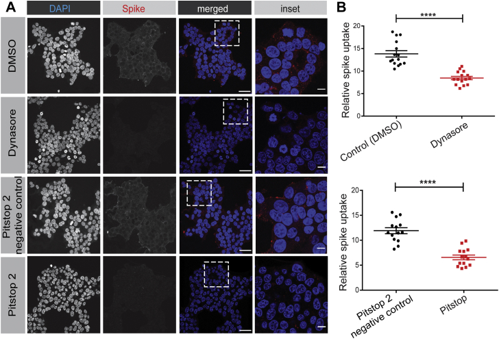
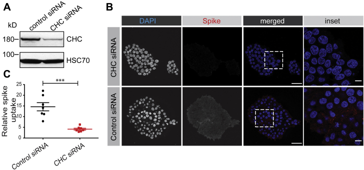
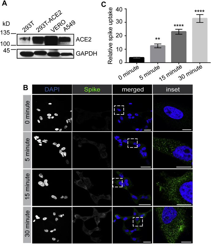
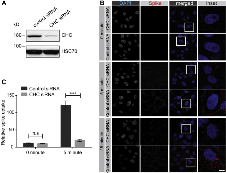
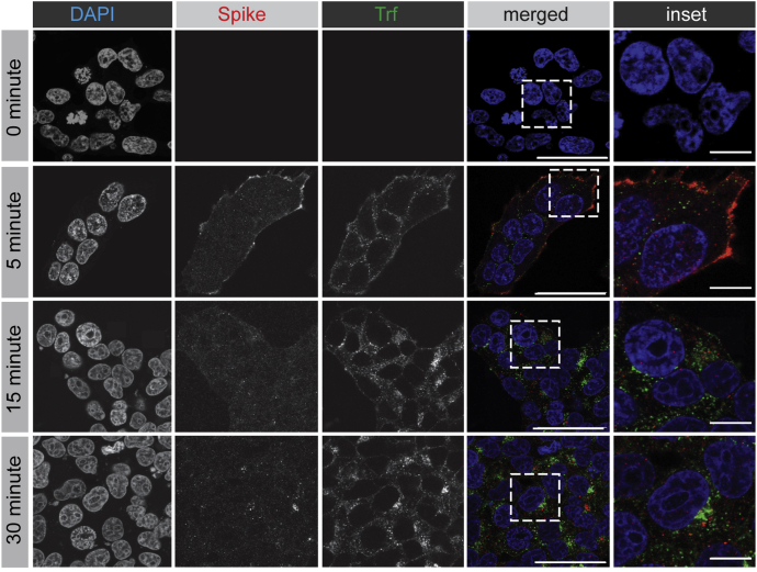
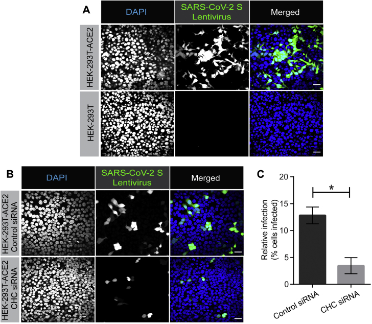
Severe acute respiratory syndrome coronavirus 2 (SARS-CoV-2) is the causative agent of COVID-19, so understanding its biology and infection mechanisms is critical to facing this major medical challenge. SARS-CoV-2 is known to use its spike glycoprotein to interact with the cell surface as a first step in the infection process. As for other coronaviruses, it is likely that SARS-CoV-2 next undergoes endocytosis, but whether or not this is required for infectivity and the precise endocytic mechanism used are unknown. Using purified spike glycoprotein and lentivirus pseudotyped with spike glycoprotein, a common model of SARS-CoV-2 infectivity, we now demonstrate that after engagement with the plasma membrane, SARS-CoV-2 undergoes rapid, clathrin-mediated endocytosis. This suggests that transfer of viral RNA to the cell cytosol occurs from the lumen of the endosomal system. Importantly, we further demonstrate that knockdown of clathrin heavy chain, which blocks clathrin-mediated endocytosis, reduces viral infectivity. These discoveries reveal that SARS-CoV-2 uses clathrin-mediated endocytosis to gain access into cells and suggests that this process is a key aspect of virus infectivity.

Keywords: COVID-19; SARS-CoV-2; clathrin; dynamin; endocytosis; infection; virus entry.


Conflict of interest statement

Conflict of interest The authors declare that they have no conflicts of interest with the contents of this article.

Figures

Figure 1
Figure 1
SARS-CoV-2 spike protein binds to the surface of HEK-293T cells expressing ACE2. A, HEK-293T cells, WT (top row of images), or stably expressing ACE2 (bottom row of images) were incubated with purified, His6-tagged spike protein and with Alexa 647–labeled transferrin (Tf) for 30 min at 4 °C. After PBS wash, the cells were fixed and stained with DAPI to reveal nuclei, with an antibody recognizing the expressed ACE2, and with an antibody recognizing the His6 epitope tag of the spike protein. Scale bars = 40 μm for the low-magnification images and 10 μm for the higher magnification inset of the merged images. B, quantification of experiments as in (A). The bar graph represents fluorescence on the plasma membrane for spike protein and Tf, from three independent experiments, mean ± SEM; unpaired t test; ∗∗∗∗p < 0.0001, n.s, not significant. C, experiments performed as in (A) except that the HEK-293T cells were briefly acid-washed before fixation. Scale bars = 40 μm for the low magnification images and 10 μm for the higher magnification inset of the composite. D, quantification of experiments as in (C). The bar graph represents fluorescence on the plasma membrane for spike protein and Tf, from three independent experiments, mean ± SEM; unpaired t test; ∗∗∗∗p < 0.0001, ∗∗p < 0.01, n.s, not significant. ACE2, angiotensin-converting enzyme 2; SARS-CoV-2, severe acute respiratory syndrome coronavirus 2.
Figure 2
Figure 2
SARS-CoV-2 spike protein enters cells in an ACE2-dependent manner. A, HEK-293T cells, WT (top row of images), or stably expressing ACE2 (bottom row of images) were incubated with purified, His6-tagged spike protein and with Alexa 647–labeled transferrin (Tf) for 30 min at 4 °C. The cells were then transferred to 37 °C for 30 min. The cells were returned to ice and after acid wash, were fixed and stained with DAPI to reveal nuclei, with an antibody recognizing the expressed ACE2, and with an antibody recognizing the His6 epitope tag of the spike protein. Scale bars = 40 μm for the low-magnification images and 10 μm for the higher magnification inset of the composite. B, quantification of experiment as in (A). The bar graph represents fluorescence on the plasma membrane for spike protein and Tf, from three independent experiments, mean ± SEM; unpaired t test; ∗∗∗p < 0.001, n.s, not significant. ACE2, angiotensin-converting enzyme 2; SARS-CoV-2, severe acute respiratory syndrome coronavirus 2.
Figure 3
Figure 3
Time course of SARS-CoV-2 spike protein entry into cells. A, HEK-293T cells stably expressing ACE2 were incubated with purified, His6-tagged spike protein for 30 min at 4 °C. The cells were then transferred to 37 °C for the indicated time periods before being returned to ice. After acid wash, cells were fixed and stained with DAPI to reveal nuclei and with antibody selectively recognizing the His6 epitope tag of the spike protein. Scale bars = 40 μm for the low-magnification images and 10 μm for the higher magnification insets on the right. B, quantification of experiments as in (A), from three independent experiments, mean ± SEM; one-way ANOVA; ∗∗∗∗p < 0.0001. ACE2, angiotensin-converting enzyme 2; SARS-CoV-2, severe acute respiratory syndrome coronavirus 2.
Figure 4
Figure 4
SARS-CoV-2 spike protein endocytosis is blocked by chemical inhibitors of clathrin-mediated endocytosis. A, HEK-293 cells stably expressing ACE2 were incubated with purified, His6-tagged spike protein for 30 min at 4 °C. The cells were then transferred to 37 °C for 30 min before being returned to ice. After acid wash, cells were fixed and stained with DAPI to reveal nuclei and with antibody selectively recognizing the His6 epitope tag of the spike protein. Inhibitors of clathrin-mediated endocytosis or their controls, as indicated, were added to the cells 30 min before the addition of spike protein. Scale bars = 40 μm for the low-magnification images and 10 μm for the higher magnification insets on the right. B, quantification of experiments performed as in (A). n = 15 for Dynasore and n = 14 for Pitstop 2 from three independent experiments, mean ± SEM; unpaired t test; ∗∗∗∗p < 0.0001. ACE2, angiotensin-converting enzyme 2; SARS-CoV-2, severe acute respiratory syndrome coronavirus 2.
Figure 5
Figure 5
SARS-CoV-2 spike protein endocytosis is reduced by CHC knockdown. A, HEK-293T cells stably expressing ACE2 were transfected with control siRNA or with an siRNA established to selectively knockdown the expression of CHC. Cell lysates were prepared and immunoblotted with the indicated antibodies. B, HEK-293T cells stably expressing ACE2, transfected with a control siRNA or an siRNA driving CHC knockdown, were incubated with purified, His6-tagged spike protein for 30 min at 4 °C. The cells were then transferred to 37 °C for 30 min before being returned to ice. After acid wash, cells were fixed and stained with DAPI to reveal nuclei and with antibody recognizing the His6 epitope tag of the spike protein. Scale bars = 40 μm for the low-magnification images and 10 μm for the higher magnification insets on the right. C, quantification of experiments performed as in (B). n = 7 from three independent experiments, mean ± SEM; unpaired t test; ∗∗∗p < 0.001. ACE2, angiotensin-converting enzyme 2; CHC, clathrin heavy chain; SARS-CoV-2, severe acute respiratory syndrome coronavirus 2.
Figure 6
Figure 6
SARS-CoV-2 spike protein is rapidly endocytosed in VERO cells. A, lysates were made from HEK-293T cells, WT or expressing ACE2, VERO cells, and A549 cells and were immunoblotted with an antibody recognizing ACE2. B, VERO cells were incubated with purified, His6-tagged spike protein for 30 min at 4 °C. The cells were then transferred to 37 °C for the indicated time periods before being returned to ice. After acid wash, cells were fixed and stained with DAPI to reveal nuclei and with antibody selectively recognizing spike protein. Scale bars = 40 μm for the low-magnification images and 10 μm for the higher magnification insets on the right. C, quantification of experiments performed as in (B) from three independent experiments, mean ± SEM; one-way ANOVA; ∗∗∗∗p < 0.0001, ∗∗p < 0.01. ACE2, angiotensin-converting enzyme 2; SARS-CoV-2, severe acute respiratory syndrome coronavirus 2.
Figure 7
Figure 7
SARS-CoV-2 spike protein endocytosis is reduced by CHC knockdown in VERO cells. A, lysates were made from VERO cells transfected with control siRNA or with an siRNA established to selectively knockdown the expression of CHC. Cell lysates were prepared and immunoblotted with the indicated antibodies. B, VERO cells, treated as in (A), were incubated with purified spike protein for 30 min at 4 °C. The cells were then transferred to 37 °C for the indicated time periods before being returned to ice. After acid wash, cells were fixed and stained with DAPI to reveal nuclei and with antibody selectively recognizing spike protein. Scale bars = 40 μm for the low-magnification images and 10 μm for the higher magnification insets on the right. C, the graph showing quantification of experiments performed as in (B). n = 15 from 3 independent experiments, mean ± SEM; unpaired t test; ∗∗∗∗p < 0.0001, n.s, not significant. ACE2, angiotensin-converting enzyme 2; CHC, clathrin heavy chain; SARS-CoV-2, severe acute respiratory syndrome coronavirus 2.
Figure 8
Figure 8
SARS-CoV-2 spike protein follows a trafficking itinerary distinct from transferrin. HEK-293T cells stably expressing ACE2 were incubated with purified, His6-tagged spike protein and Trf-Alexa647 for 30 min at 4 °C. The cells were then transferred to 37 °C for the indicated time periods before being returned to ice. After PBS wash, cells were fixed and stained with DAPI to reveal nuclei and with antibody selectively recognizing the His6 epitope tag of the spike protein. Scale bars = 40 μm for the low-magnification images and 10 μm for the higher magnification insets on the right. ACE2, angiotensin-converting enzyme 2; SARS-CoV-2, severe acute respiratory syndrome coronavirus 2.
Figure 9
Figure 9
Lentivirus pseudotyped with SARS-CoV-2 spike glycoprotein requires CHC for infectivity. A, HEK-293T cells stably expressing ACE2 or HEK-293T cells were incubated with lentivirus pseudotyped with the SARS-CoV-2 spike glycoprotein for 12 h. Cells were then fixed and stained with DAPI to reveal nuclei. Cells were also imaged for GFP, driven from the pseudovirus. Scale bars = 40 μm. B, HEK-293T cells stably expressing ACE2 were transfected with control siRNA or with an siRNA established to selectively knockdown the expression of CHC. The cells were incubated with lentivirus pseudotyped with the SARS-CoV-2 spike glycoprotein for 12 h. Cells were then fixed and stained with DAPI to reveal nuclei. Cells were also imaged for GFP, driven from the pseudovirus. Scale bars = 40 μm. C, the graph showing quantification of SARS-CoV-2 pseudovirus infection from experiments as in (B). n = 3 from three independent experiments, mean ± SEM; unpaired t test, ∗p < 0.05. ACE2, angiotensin-converting enzyme 2; CHC, clathrin heavy chain; SARS-CoV-2, severe acute respiratory syndrome coronavirus 2.

References

    1. Zaki A.M., van Boheemen S., Bestebroer T.M., Osterhaus A.D., Fouchier R.A. Isolation of a novel coronavirus from a man with pneumonia in Saudi Arabia. N. Engl. J. Med. 2012;367:1814–1820. - PubMed
    1. Drosten C., Günther S., Preiser W., van der Werf S., Brodt H.R., Becker S., Rabenau H., Panning M., Kolesnikova L., Fouchier R.A., Berger A., Burguière A.M., Cinatl J., Eickmann M., Escriou N. Identification of a novel coronavirus in patients with severe acute respiratory syndrome. N. Engl. J. Med. 2003;348:1967–1976. - PubMed
    1. Ksiazek T.G., Erdman D., Goldsmith C.S., Zaki S.R., Peret T., Emery S., Tong S., Urbani C., Comer J.A., Lim W., Rollin P.E., Dowell S.F., Ling A.E., Humphrey C.D., Shieh W.J. A novel coronavirus associated with severe acute respiratory syndrome. N. Engl. J. Med. 2003;348:1953–1966. - PubMed
    1. Huang C., Wang Y., Li X., Ren L., Zhao J., Hu Y., Zhang L., Fan G., Xu J., Gu X. Clinical features of patients infected with 2019 novel coronavirus in Wuhan, China. Lancet. 2020;395:497–506. - PMC - PubMed
    1. Zhu N., Zhang D., Wang W., Li X., Yang B., Song J., Zhao X., Huang B., Shi W., Lu R. A Novel coronavirus from patients with pneumonia in China. N. Engl. J. Med. 2020;382:727–733. - PMC - PubMed

Publication types

MeSH terms