. 2021 Jan 17;14(2):436.
doi: 10.3390/ma14020436.

Characterization of Properties, In Vitro and In Vivo Evaluation of Calcium Phosphate/Amino Acid Cements for Treatment of Osteochondral Defects

Affiliations

Characterization of Properties, In Vitro and In Vivo Evaluation of Calcium Phosphate/Amino Acid Cements for Treatment of Osteochondral Defects

Lubomir Medvecky et al. Materials (Basel). .

Abstract

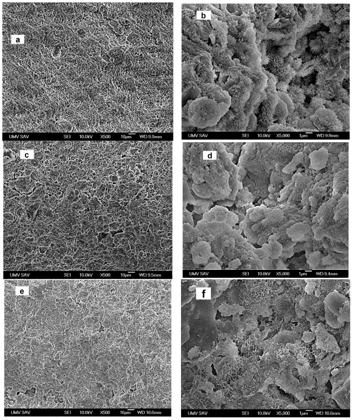
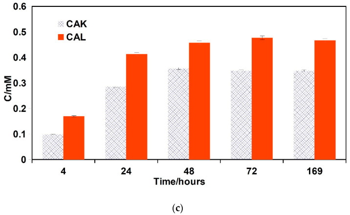
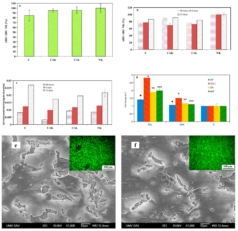
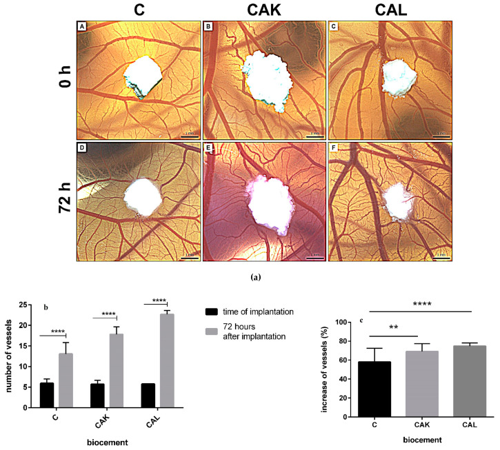
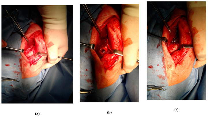
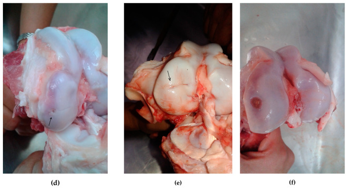
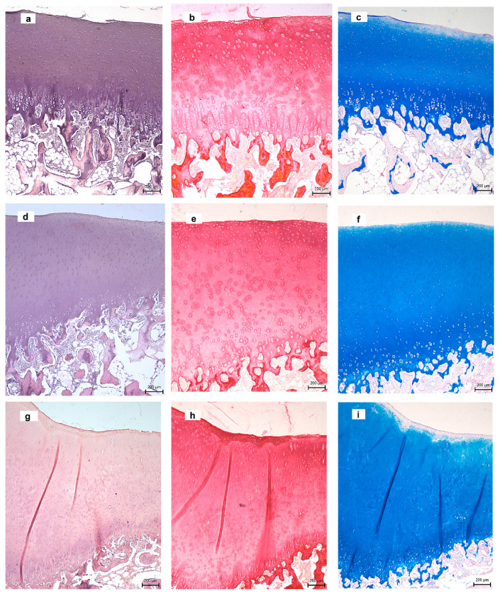
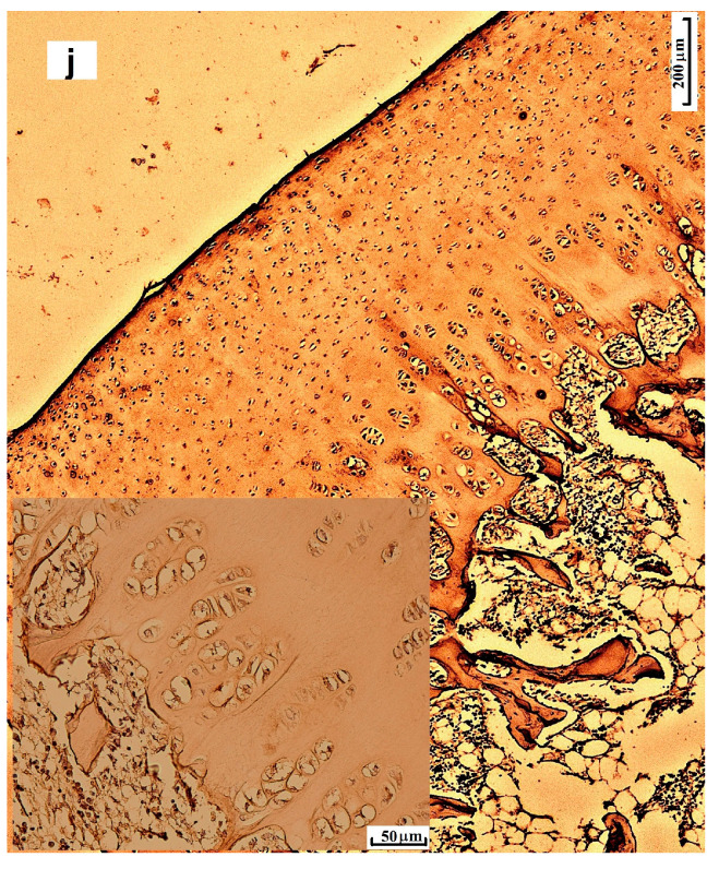
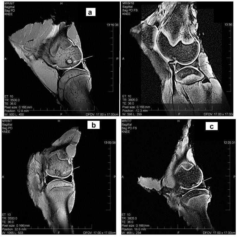
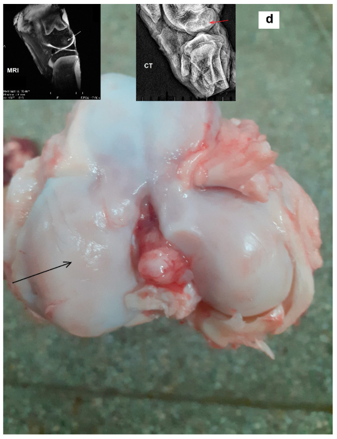
Novel calcium phosphate cements containing a mixture of four amino acids, glycine, proline, hydroxyproline and either lysine or arginine (CAL, CAK) were characterized and used for treatment of artificial osteochondral defects in knee. It was hypothesized that an enhanced concentration of extracellular collagen amino acids (in complex mixture), in connection with bone cement in defect sites, would support the healing of osteochondral defects with successful formation of hyaline cartilage and subchondral bone. Calcium phosphate cement mixtures were prepared by in situ reaction in a planetary ball mill at aseptic conditions and characterized. It was verified that about 30-60% of amino acids remained adsorbed on hydroxyapatite particles in cements and the addition of amino acids caused around 60% reduction in compressive strength and refinement of hydroxyapatite particles in their microstructure. The significant over-expression of osteogenic genes after the culture of osteoblasts was demonstrated in the cement extracts containing lysine and compared with other cements. The cement pastes were inserted into artificial osteochondral defects in the medial femoral condyle of pigs and, after 3 months post-surgery, tissues were analyzed macroscopically, histologically, immunohistochemically using MRI and X-ray methods. Analysis clearly showed the excellent healing process of artificial osteochondral defects in pigs after treatment with CAL and CAK cements without any inflammation, as well as formation of subchondral bone and hyaline cartilage morphologically and structurally identical to the original tissues. Good integration of the hyaline neocartilage with the surrounding tissue, as well as perfect interconnection between the neocartilage and new subchondral bone tissue, was demonstrated. Tissues were stable after 12 months' healing.

Keywords: amino acid; calcium phosphate cement; hyaline cartilage; osteochondral defect; pig model.


Conflict of interest statement

The authors declare no conflict of interest.

Figures

Figure 1
Figure 1
XRD patterns (a) (M-monetite, ○-HAP, T-TTCP) and FTIR spectra (b) of cement mixtures and cements after 7 days soaking at 37 °C in SBF (a–CAK mix, b–CAL mix, c–CAK cem, d–CAL cem).
Figure 2
Figure 2
Microstructures of cements after 7 days setting in SBF ((a,b)–C; (c,d)–CAK; (e,f)–CAL).
Figure 3
Figure 3
Release of amino acids (CAK—(a), CAL—(b)) and calcium (c) from cements to 0.9% NaCl solution at 37 °C.
Figure 3
Figure 3
Release of amino acids (CAK—(a), CAL—(b)) and calcium (c) from cements to 0.9% NaCl solution at 37 °C.
Figure 4
Figure 4
Viability of osteoblasts cultured in A cement extracts for 24 h (a), B cement extracts during the prolonged cultivation period of up to 15 days (b), ALP activity of osteoblasts cultured in B cement extracts (c), relative gene expression of COL1, ON, OP; ALP in osteoblasts cultured for 7 days in B extracts of CAL (statistically significant differences, p < 0.05) (d); SEM micrographs and live/dead staining of osteoblasts cultured for 9 days in osteogenic medium on CAK (e) and CAL (f) hardened cements.
Figure 5
Figure 5
Quail chorioallantoic membrane (CAM) assay (a): representative photographs with the growing of vessels toward the biomaterials in the time of implantation (0 h; A–C) and 72 h after implantation (D–F), scale bar = 1 mm; quantification of quail CAM vessels at 0 h and 72 h after implantation of biocements (b). Results are summarized in the graph as the vascular index for each tested biomaterial. The graph shows data presented as mean ± standard deviation of three independent experiments (**** p < 0.0001 per each biomaterial); angiogenic activity of biocements 72 h after their implantation (** p < 0.01; **** p < 0.0001) (c).
Figure 6
Figure 6
The operating procedure (top). Left porcine stifle joint, medial condyle (a), created defect (Φ 10 mm) in the articular cartilage and subchondral bone (b), drill hole was filled by biocement (c). Bottom: macroscopic evaluation of the osteochondral defect healing with biocements (d) CAL, (e) CAK, (f) C.
Figure 6
Figure 6
The operating procedure (top). Left porcine stifle joint, medial condyle (a), created defect (Φ 10 mm) in the articular cartilage and subchondral bone (b), drill hole was filled by biocement (c). Bottom: macroscopic evaluation of the osteochondral defect healing with biocements (d) CAL, (e) CAK, (f) C.
Figure 7
Figure 7
Histological analysis of tissues in defects after 3 months’ healing ((a–c) CAL; (d–f) CAK; (g–i) C) stained with hematoxylin & eosin (a,d,g)), Picrosirius red (b,e,h), Alcian blue (c,f,i), collagen II in CAL (j). (Arrows—defect sites.)
Figure 7
Figure 7
Histological analysis of tissues in defects after 3 months’ healing ((a–c) CAL; (d–f) CAK; (g–i) C) stained with hematoxylin & eosin (a,d,g)), Picrosirius red (b,e,h), Alcian blue (c,f,i), collagen II in CAL (j). (Arrows—defect sites.)
Figure 8
Figure 8
MRI assessment of cartilage defects after 3 months’ healing, the sagittal plane reconstruction of the articular cartilage defect examined, on the left, porcine stifle joint in a 1,2 T high-field MRI setting ((a) CAL, (b) CAK, (c) C); macroscopic and MRI assessment of cartilage defect treated with CAL after 12 months’ healing illustrating the macroscopic picture of the exposed stifle joint after euthanasia (d) (sagittal plane reconstruction of the articular cartilage defect examined in a 1,2 T high-field MRI setting and lateral projection, on the left, porcine stifle joint, the medial condyle examined by X-ray in a Philips Digital Diagnost). (Arrows—defect sites.)
Figure 8
Figure 8
MRI assessment of cartilage defects after 3 months’ healing, the sagittal plane reconstruction of the articular cartilage defect examined, on the left, porcine stifle joint in a 1,2 T high-field MRI setting ((a) CAL, (b) CAK, (c) C); macroscopic and MRI assessment of cartilage defect treated with CAL after 12 months’ healing illustrating the macroscopic picture of the exposed stifle joint after euthanasia (d) (sagittal plane reconstruction of the articular cartilage defect examined in a 1,2 T high-field MRI setting and lateral projection, on the left, porcine stifle joint, the medial condyle examined by X-ray in a Philips Digital Diagnost). (Arrows—defect sites.)

References

    1. Conaghan P.G., Kloppenburg M., Schett G., Bijlsma J.W.J., EULAR osteoarthritis ad hoc committee Osteoarthritis research priorities: A report from a EULAR ad hoc expert committee. Ann. Rheum. Dis. 2014;73:1442–1445. doi: 10.1136/annrheumdis-2013-204660. - DOI - PubMed
    1. Zhang Y., Jordan J.M. Epidemiology of osteoarthritis. Clin. Geriatr. Med. 2010;26:355–369. doi: 10.1016/j.cger.2010.03.001. - DOI - PMC - PubMed
    1. Nukavarapu S.P., Dorcemus D.L. Osteochondral tissue engineering: Current strategies and challenges. Biotechnol. Adv. 2013;31:706–721. doi: 10.1016/j.biotechadv.2012.11.004. - DOI - PubMed
    1. Cole B.J., Harris J.D. Biologic Knee Reconstruction: A Surgeon’s Guide. 1st ed. SLACK, Inc.; Thorofare, NJ, USA: 2015. ISBN-13: 978-1617118166; ISBN-10: 1617118168.
    1. Puppi D., Chiellini F., Piras A.M., Chiellini E. Polymeric materials for bone and cartilage repair. Prog. Polym. Sci. 2010;35:403–440. doi: 10.1016/j.progpolymsci.2010.01.006. - DOI

LinkOut - more resources