. 2021 Jan 17;9(1):189.
doi: 10.3390/microorganisms9010189.

Microbiome Signatures in a Fast- and Slow-Progressing Gastric Cancer Murine Model and Their Contribution to Gastric Carcinogenesis

Affiliations

Microbiome Signatures in a Fast- and Slow-Progressing Gastric Cancer Murine Model and Their Contribution to Gastric Carcinogenesis

Prerna Bali et al. Microorganisms. .

Abstract

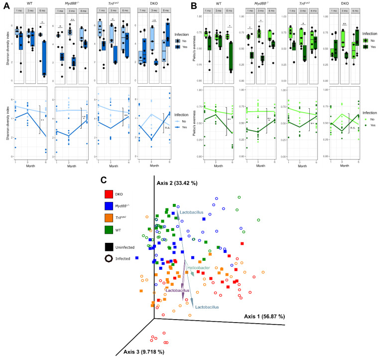
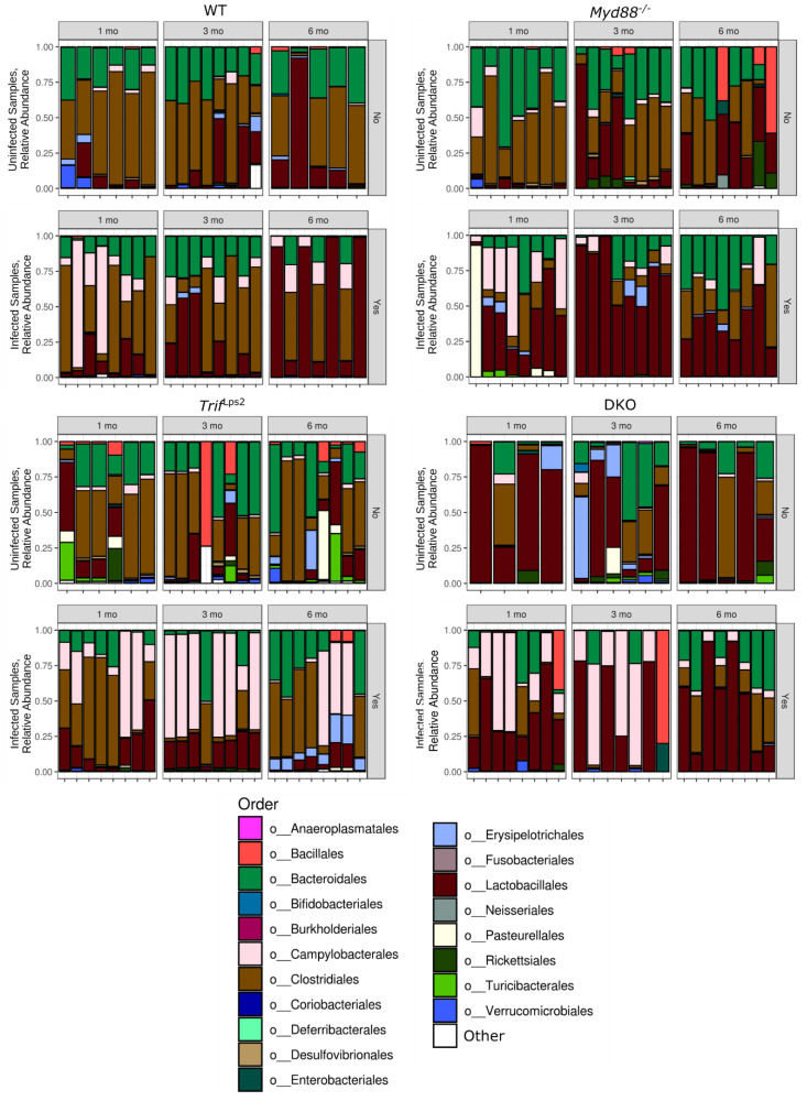
Gastric cancer is the third most common cause of death from cancer in the world and infection with Helicobacter pylori (H. pylori) is the main cause of gastric cancer. In addition to Helicobacter infection, the overall stomach microbiota has recently emerged as a potential factor in gastric cancer progression. Previously we had established that mice deficient in myeloid differentiation primary response gene 88 (MyD88, Myd88-/- ) rapidly progressed to neoplasia when infected with H. felis. Thus, in order to assess the role of the microbiota in this fast-progressing gastric cancer model we investigated changes of the gastric microbiome in mice with different genotypic backgrounds: wild type (WT), MyD88-deficient (Myd88-/- ), mice deficient in the Toll/interleukin-1 receptor (TIR) domain-containing adaptor-inducing interferon-β (TRIF, Trif Lps2), and MyD88- and TRIF-deficient (Myd88-/- /Trif Lps2, double knockout (DKO)) mice. We compared changes in alpha diversity, beta diversity, relative abundance, and log-fold differential of relative abundance ratios in uninfected and Helicobacter infected mice and studied their correlations with disease progression to gastric cancer in situ. We observed an overall reduction in microbial diversity post-infection with H. felis across all genotypes. Campylobacterales were observed in all infected mice, with marked reduction in abundance at 3 and 6 months in Myd88-/- mice. A sharp increase in Lactobacillales in infected Myd88-/- and DKO mice at 3 and 6 months was observed as compared to Trif Lps2 and WT mice, hinting at a possible role of these bacteria in gastric cancer progression. This was further reinforced upon comparison of Lactobacillales log-fold differentials with histological data, indicating that Lactobacillales are closely associated with Helicobacter infection and gastric cancer progression. Our study suggests that differences in genotypes could influence the stomach microbiome and make it more susceptible to the development of gastric cancer upon Helicobacter infection. Additionally, increase in Lactobacillales could contribute to faster development of gastric cancer and might serve as a potential biomarker for the fast progressing form of gastric cancer.

Keywords: Helicobacter; Lactobacillales; MyD88; TRIF; gastric cancer; microbiome.


Conflict of interest statement

The authors state they have no conflict of interest to declare.

Figures

Figure 1
Figure 1
Changes in microbial diversity in the stomach across four different genotypes, with infection and time. Shannon diversity index (A) and Pielou’s evenness (B) values for each gastric community, divided by genotype and month. Top and bottom plots represent the same data, alpha diversity values, with lines on the bottom plots denoting average values for infected and uninfected communities over time. Statistical significance on bottom plots refers to differences between infected and uninfected, months 1–6 combined. (C) Principal component analysis of robust Aitchison distance values between communities, months 1–6 combined. Biplot arrows indicate operational taxonomic units (OTUs) driving separation between samples, with arrows labeled with the genus of the OTU. Arrows and genus labels are matched by color. All diversity metrics were calculated using QIIME2. Statistical significance determined by Student’s t-test for alpha diversity and permutational multivariate analysis of variance (PERMANOVA) with Benjamini-Hochberg FDR correction for beta diversity (* p < 0.05, ** p < 0.01, *** p < 0.005).
Figure 2
Figure 2
Relative abundance of different phyla across four genotypes. Relative abundance of individual phyla from WT, Myd88−/−, TrifLps2, and double knockout (DKO) genotypes. The top eight phyla are shown in the legend. Samples are grouped into Helicobacter-infected and non-Helicobacter-infected and divided by time point. Sequencing data were processed in QIIME2, then plotted in PhyloSeq.
Figure 3
Figure 3
Relative abundance of different orders across four genotypes. Relative abundance of the top 15 orders from WT, Myd88−/−, TrifLps2, and DKO genotypes. Remaining phyla are grouped into “Other”. Samples are grouped into Helicobacter-infected and non-Helicobacter-infected and divided by time point. Sequencing data were processed in QIIME2, then plotted in PhyloSeq.
Figure 4
Figure 4
Log ratios between relevant orders across four genotypes. Log ratios of the Lactobacillales/Rickettsiales (A,B) and Clostridiales/Rickettsiales (C,D) relative abundance ratios between samples, months 1–6 combined. (A,C) show ratios by infection status, all genotypes combined. (B,D) show ratios by genotype and infection status. Log ratios were calculated and processed using Songbird and Qurro. Statistical significance was determined by analysis of variance (ANOVA) ** p < 0.01, *** p < 0.005).
Figure 5
Figure 5
Histopathological scoring for mucous metaplasia. Following infection with H. felis for 1 month (A), 3 months (B) and 6 months (C), H&E-stained stomach sections from each mouse (WT and Myd88−/−, TrifLps2and DKO) were evaluated for indications of pathology. Mucous metaplasia was scored by a blinded comparative pathologist according to the criteria described in Materials and Methods. A p value of 0.05 was considered statistically significant. (A) 1month post infection, n = 14 for WT, n = 15 for Myd88−/−, n = 14 for TrifLps2, and n = 12 for DKO mice; (B) 3 months post infection, n = 16 for WT, n = 16 for Myd88−/−, n = 16 for TrifLps2, n = 13 for DKO mice; (C) 6 months post infection, n = 12 for WT, n = 16 for Myd88−/−, n = 16 for TrifLps2, n = 12 for DKO mice. Statistical significance was determined by Mann-Whitney test, *** p < 0.005).
Figure 6
Figure 6
Predictive relationship between Lactobacillales and mucous metaplasia from Helicobacter infection. (A,B) Ordinal logistic regression analysis of log ratios of Lactobacillales/Rickettsiales (A), and Clostridiales/Rickettsiales (B) Relative abundance and gastric mucous histology score. The black circle marks the average of each category. Ordinal logistic regression was calculated using the polr command in R (*** p < 0.001). (CE) ROC curve for log ratios of Campylobacterales, Lactobacillales, and Clostridiales to Rickettsiales. The blue line represents the performance of each ratio log fold-differential in predicting Helicobacter infection. The red line represents the result expected for a metric with a 50% chance of predicting infection. The area under the curve (AUC) value refers to the area under the blue line. Receiver operating characteristic (ROC) plots were constructed in Prism7 using log fold-differentials from Songbird and Qurro.

References

    1. World Health Organization Cancer. WHO. [(accessed on 12 September 2018)]; Available online: https://www.who.int/news-room/fact-sheets/detail/cancer.
    1. Schistosomes, liver flukes and Helicobacter pylori. IARC Working Group on the Evaluation of Carcinogenic Risks to Humans. Lyon, 7–14 June 1994. [(accessed on 15 January 2021)];IARC Monogr. Eval. Carcinog. Risks Hum. 1994 61:1–241. Available online: https://www.ncbi.nlm.nih.gov/books/NBK487782/ - PMC - PubMed
    1. Noto J.M., Peek R.M., Jr. The gastric microbiome, its interaction with Helicobacter pylori, and its potential role in the progression to stomach cancer. PLoS Pathog. 2017;13:e1006573. doi: 10.1371/journal.ppat.1006573. - DOI - PMC - PubMed
    1. Aziz F., Chakarobaty A., Liu K., Yoshitomi H., Li X., Monts J., Xu G., Li Y., Bai R., Bode A.M., et al. Gastric tumorigenesis induced either by Helicobacter pylori infection or chronic alcohol consumption through IL-10 inhibition. Res. Sq. 2020 doi: 10.21203/rs.2.23065/v1. - DOI
    1. Majmudar K., Golemi I., Tafur A.J., Toro J.D., Visonà A., Falgá C., Sahuquillo J.C., Lorente M.A., Tufano A., Weinberg I., et al. RIETE Investigators. Outcomes after venous thromboembolism in patients with gastric cancer: Analysis of the RIETE Registry. Vasc. Med. 2020;25:210–217. doi: 10.1177/1358863X19893432. - DOI - PubMed