. 2021 Jan 17;11(1):62.
doi: 10.3390/membranes11010062.

In Vitro Evaluation of Polihexanide, Octenidine and NaClO/HClO-Based Antiseptics against Biofilm Formed by Wound Pathogens

Affiliations

In Vitro Evaluation of Polihexanide, Octenidine and NaClO/HClO-Based Antiseptics against Biofilm Formed by Wound Pathogens

Grzegorz Krasowski et al. Membranes (Basel). .

Abstract

Chronic wounds complicated with biofilm formed by pathogens remain one of the most significant challenges of contemporary medicine. The application of topical antiseptic solutions against wound biofilm has been gaining increasing interest among clinical practitioners and scientific researchers. This paper compares the activity of polyhexanide-, octenidine- and hypochlorite/hypochlorous acid-based antiseptics against biofilm formed by clinical strains of Candida albicans, Staphylococcus aureus and Pseudomonas aeruginosa. The analyses included both standard techniques utilizing polystyrene plates and self-designed biocellulose-based models in which a biofilm formed by pathogens was formed on an elastic, fibrinous surface covered with a fibroblast layer. The obtained results show high antibiofilm activity of polihexanide- and octenidine-based antiseptics and lack or weak antibiofilm activity of hypochlorite-based antiseptic of total chlorine content equal to 80 parts per million. The data presented in this paper indicate that polihexanide- or octenidine-based antiseptics are highly useful in the treatment of biofilm, while hypochlorite-based antiseptics with low chlorine content may be applied for wound rinsing but not when specific antibiofilm activity is required.

Keywords: hypochlorous acid; octenidine; polihexanide; sodium hypochlorite; wound biofilm.


Conflict of interest statement

The authors declare no conflict of interest. The funders had no role in the design of the study; in the collection, analyses, or interpretation of data; in the writing of the manuscript, or in the decision to publish the results.

Figures

Figure 1
Figure 1
Minimal biofilm eradication concentration (MBEC) of P and O antiseptics against the tested strains (n = 9) of C. albicans, S. aureus, P. aeruginosa. In the case of the M antiseptic, the MBEC value for all pathogenic biofilms tested was higher than the maximum concentration (i.e., 50% of the volume of the product provided) which could be applied according to this methodology. The asterisks (*) in the bottom figure show a statistically significant difference between the ability of the O- antiseptic to eradicate P. aeruginosa biofilms vs. C. albicans and S. aureus biofilms; p < 0.05). P, O—polihexanide, octenidine dihydrochloride-based antiseptic, respectively.
Figure 2
Figure 2
Presentation of cellulose-based biofilm model. (A) native cellulose-carrier (SEM, magn. 5000×); (B) cellulose carrier covered with fibroblasts (SEM, magn. 2500×); (C) staphylococcal (PRT9) biofilm formed on fibroblast-containing cellulose carrier visualized with confocal microscopy. Big red (dead) and green (live) oval shapes—fibroblasts; smaller green dots—staphylococcal cells. The figure shows a side view projection of the cellulose carrier (not visible) covered with a fibroblast layer and bacteria. A high share of dead fibroblasts (red oval shapes) during 24 h of co-culture with staphylococci is worth noting.
Figure 3
Figure 3
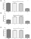
Eradication of (A) C. albicans; (B) S. aureus, (C) P. aeruginosa biofilm from fibriblast-covered cellulose carrier by P, O and M antiseptics. P, O, M—polihexanide, octenidine dihydrochloride, NaClO/HClO-based antiseptics, respectively. H2O2 of concentration 30% was used as a control of microbial killing, while the number of biofilm-forming cells immersed in saline were considered 100% of potential microbial viability. The asterisks (*) represent statistical significance (p < 0.05) between individual antiseptics’ activity.
Figure 4
Figure 4
Live/dead images of biofilm-forming cells forming on a cellulose carrier subjected to the antiseptics for 1 h. (A) untreated biofilm (no antimicrobial solution); (B) 30% H2O2, biofilm treated with 30% hydrogen peroxide, i.e., compound of strong antimicrobial activity; (C) biofilm treated with P antiseptic; (D) biofilm treated with O antiseptic; (E) biofilm treated with M antiseptic. Green—live cells, red/orange—dead cells. The white bars in the right lower part of every picture are of 50 µm of longitude. Please also refer to supplementary data Figures S6 and S7 where pictures of bigger size with characters and typical points are presented.

References

    1. Kadam S., Shai S., Shahane A., Kaushik K.S. Recent Advances in Non-Conventional Antimicrobial Approaches for Chronic Wound Biofilms: Have We Found the ‘Chink in the Armor’? [(accessed on 2 December 2020)];Biomedicines. 2019 7:35. doi: 10.3390/biomedicines7020035. Available online: https://www.mdpi.com/2227-9059/7/2/35#cite. - DOI - PMC - PubMed
    1. Lappin-Scott H., Burton S., Stoodley P. Revealing a world of biofilms—The pioneering research of Bill Costerton. Nat. Rev. Genet. 2014;12:781–787. doi: 10.1038/nrmicro3343. - DOI - PubMed
    1. Dongari-Bagtzoglou A. Pathogenesis of mucosal biofilm infections: Challenges and progress. Expert Rev. Anti-infect. Ther. 2008;6:201–208. doi: 10.1586/14787210.6.2.201. - DOI - PMC - PubMed
    1. Muhammad M.H., Idris A.L., Fan X., Guo Y., Yu Y., Jin X., Qiu J., Guan X., Huang T. Beyond Risk: Bacterial Biofilms and Their Regulating Approaches. Front. Microbiol. 2020;11:928. doi: 10.3389/fmicb.2020.00928. - DOI - PMC - PubMed
    1. Mikulskis P., Hook A.L., Dundas A.A., Irvine D.J., Sanni O., Anderson D.G., Langer R., Alexander M.R., Williams P., Winkler D.A. Prediction of Broad-Spectrum Pathogen Attachment to Coating Materials for Biomedical Devices. ACS Appl. Mater. Interfaces. 2018;10:139–149. doi: 10.1021/acsami.7b14197. - DOI - PMC - PubMed

LinkOut - more resources