Review
. 2021 Jan 18;22(2):897.
doi: 10.3390/ijms22020897.

Molecular Changes Underlying Hypertrophic Scarring Following Burns Involve Specific Deregulations at All Wound Healing Stages (Inflammation, Proliferation and Maturation)

Affiliations
Review

Molecular Changes Underlying Hypertrophic Scarring Following Burns Involve Specific Deregulations at All Wound Healing Stages (Inflammation, Proliferation and Maturation)

Matúš Čoma et al. Int J Mol Sci. .

Abstract

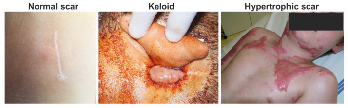
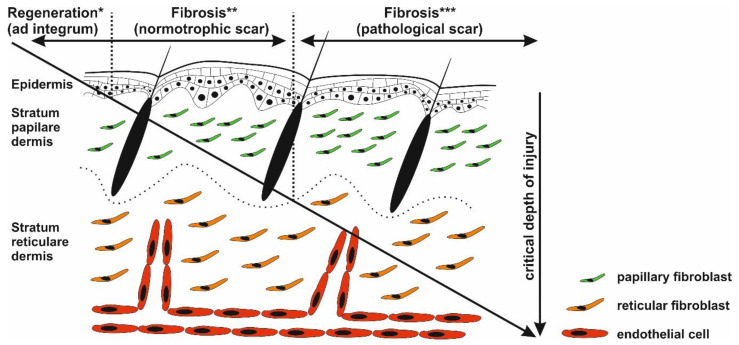
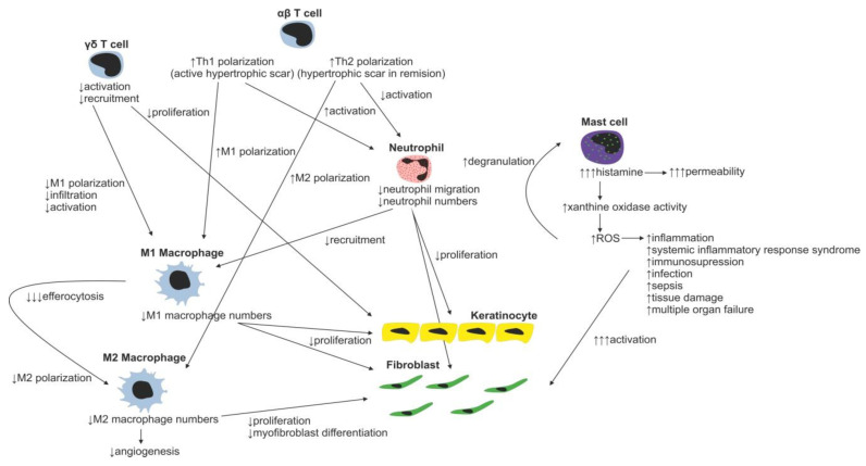
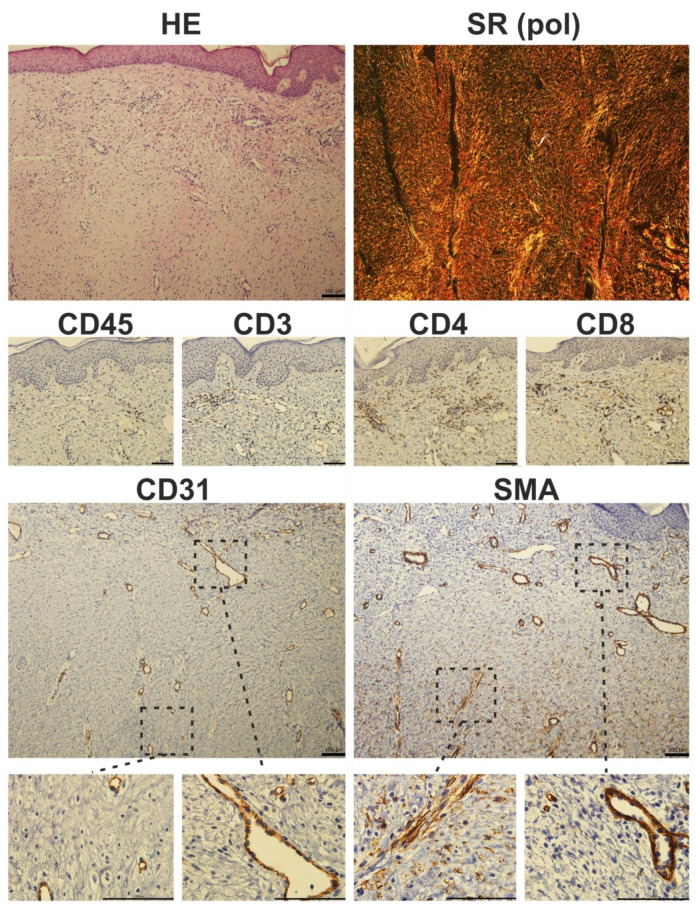
Excessive connective tissue accumulation, a hallmark of hypertrophic scaring, results in progressive deterioration of the structure and function of organs. It can also be seen during tumor growth and other fibroproliferative disorders. These processes result from a wide spectrum of cross-talks between mesenchymal, epithelial and inflammatory/immune cells that have not yet been fully understood. In the present review, we aimed to describe the molecular features of fibroblasts and their interactions with immune and epithelial cells and extracellular matrix. We also compared different types of fibroblasts and their roles in skin repair and regeneration following burn injury. In summary, here we briefly review molecular changes underlying hypertrophic scarring following burns throughout all basic wound healing stages, i.e. during inflammation, proliferation and maturation.

Keywords: burn; cell interaction; pathological scar; skin; stem cell; wound healing.


Conflict of interest statement

The authors declare no conflict of interest.

Figures

Figure 1
Figure 1
Clinical photos of normal, keloid and hypertrophic scars: normal fine line scar following a surgical wound (left); keloid scar, which extends beyond the original wound (middle); and hypertrophic scar, which does not extend beyond the initial site of the skin lesion associated with burn injury (right).
Figure 2
Figure 2
Critical depth of injury. Only the epidermis has the capability to fully regenerate (*), whereas healing of the injured dermis results in scar formation (** and ***). Injury targeting only papillary dermis ends with normotrophic scar formation (*). Deeper wounds (reticular parts of the dermis) with prolonged inflammatory response may activate biological pathways resulting in pathological scarring (***).
Figure 3
Figure 3
Immune cell regulation of normal wound healing.
Figure 4
Figure 4
Changes in immune cell regulation of wound healing following burn injury.
Figure 5
Figure 5
Primary cultures of human dermal fibroblasts isolated from matured scar (Scar), granulation tissue (GT) and split-thickness skin graft (STSG). Cultures were stained for α-smooth muscle actin (SMA, red signal), fibronectin (Fibr, green signal) and nucleus (DAPI, blue signal) (scale bar 100 µm).
Figure 6
Figure 6
Histological photomicrograph of a hypertrophic scar stained with hematoxylin-eosin (HE) and Sirius red (SR) observed under polarized light (pol). Immunohistochemical staining of a hypertrophic scar. Prolonged inflammation results in the presence of CD45-positive cells in fibrotic tissue. CD3 staining showed characteristic T-cell islands, which were more pronounced after selective staining of CD4 and CD8-expressing T-cell subpopulations. The presence of myofibroblasts is restricted to deeper parts of the scar: in detail, α-smooth muscle actin (SMA) positivity was seen in luminized vessels (also positive for endothelial marker CD31), whereas CD31-negative and SMA-expressing cells (myofibroblasts) were located rather in the deeper parts of the scars (scale 100 µm).

References

    1. Bayat A., McGrouther D.A., Ferguson M.W. Skin scarring. BMJ. 2003;326:88–92. doi: 10.1136/bmj.326.7380.88. - DOI - PMC - PubMed
    1. Tsao S.S., Dover J.S., Arndt K.A., Kaminer M.S. Scar management: Keloid, hypertrophic, atrophic, and acne scars. Semin. Cutan. Med. Surg. 2002;21:46–75. doi: 10.1053/sder.2002.31153. - DOI - PubMed
    1. Bell L., McAdams T., Morgan R., Parshley P.F., Pike R.C., Riggs P., Carpenter J.E. Pruritus in burns: A descriptive study. J. Burn Care Rehabil. 1988;9:305–308. - PubMed
    1. Robert R., Blakeney P., Villarreal C., Meyer W.J., 3rd Anxiety: Current practices in assessment and treatment of anxiety of burn patients. Burns. 2000;26:549–552. doi: 10.1016/S0305-4179(00)00016-4. - DOI - PubMed
    1. Taal L.A., Faber A.W. Posttraumatic stress and maladjustment among adult burn survivors 1-2 years postburn. Burns. 1998;24:285–292. doi: 10.1016/S0305-4179(98)00030-8. - DOI - PubMed

LinkOut - more resources