Review
. 2021 Jan 18;22(2):904.
doi: 10.3390/ijms22020904.

Amyotrophic Lateral Sclerosis Genes in Drosophila melanogaster

Affiliations
Review

Amyotrophic Lateral Sclerosis Genes in Drosophila melanogaster

Sophie Layalle et al. Int J Mol Sci. .

Abstract

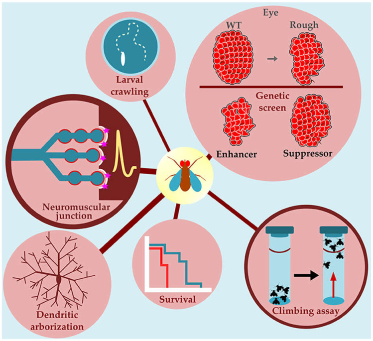
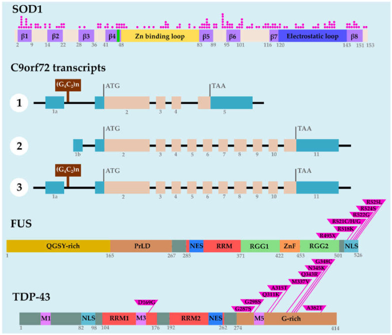
Amyotrophic lateral sclerosis (ALS) is a devastating adult-onset neurodegenerative disease characterized by the progressive degeneration of upper and lower motoneurons. Most ALS cases are sporadic but approximately 10% of ALS cases are due to inherited mutations in identified genes. ALS-causing mutations were identified in over 30 genes with superoxide dismutase-1 (SOD1), chromosome 9 open reading frame 72 (C9orf72), fused in sarcoma (FUS), and TAR DNA-binding protein (TARDBP, encoding TDP-43) being the most frequent. In the last few decades, Drosophila melanogaster emerged as a versatile model for studying neurodegenerative diseases, including ALS. In this review, we describe the different Drosophila ALS models that have been successfully used to decipher the cellular and molecular pathways associated with SOD1, C9orf72, FUS, and TDP-43. The study of the known fruit fly orthologs of these ALS-related genes yielded significant insights into cellular mechanisms and physiological functions. Moreover, genetic screening in tissue-specific gain-of-function mutants that mimic ALS-associated phenotypes identified disease-modifying genes. Here, we propose a comprehensive review on the Drosophila research focused on four ALS-linked genes that has revealed novel pathogenic mechanisms and identified potential therapeutic targets for future therapy.

Keywords: C9orf72; Drosophila melanogaster; FUS; SOD1; TDP-43; amyotrophic lateral sclerosis.


Conflict of interest statement

The authors declare no conflict of interest.

Figures

Figure 1
Figure 1
Drosophila genetic tools. The Gal4 system introduced in Drosophila allows transgene ectopic expression with spatiotemporal control. The upstream activating sequence (UAS)/Gal4 is a bipartite system. One transgenic line expresses the Gal4 transcription factor from yeast in a tissue-specific manner using an endogenous promoter or an enhancer sequence. The other line carries the transgene under the control of the UAS (upstream activating sequence). In the progeny of the cross, the UAS is bound by Gal4 protein and the transcription of the gene of interest starts. The temporal and regional gene expression targeting (TARGET) method allows a more flexible temporal control of the Gal4 system. A temperature-sensitive version of Gal80 protein (Gal80TS) is expressed ubiquitously under the control of the Tubulin promoter. At permissive temperature (18 °C), Gal80TS bound to Gal4 and prevents the starting of the transcription. A temperature shift to 29 °C relieves the transcriptional repression and permits the transcription of the gene of interest. The GeneSwitch system is based on hormone-inducible Gal4 and allows a temporal regulation of transgene expression. Gal4 is fused to a progesterone receptor, and the addition of hormone or synthetic ligand (RU486) in the feeding of the fly (adults or larvae) activates the transcription of the gene of interest. This system is reversible as the removal of ligand silent the system. All these genetic tools are routinely used in laboratory to precisely control transgenes expression not only in a specific tissue but also in a precise temporal manner.
Figure 2
Figure 2
Drosophila is a model to study neurodegenerative diseases. Several neurodegenerative diseases, including amyotrophic lateral sclerosis (ALS), have been modeled in Drosophila via transgenic expression of wildtype or mutated human proteins. The toolkit available at Drosophila allows an in-depth study of the neurodegenerative mechanisms associated with these diseases. Several behavioral tasks, such as larval crawling and climbing assays, allow monitoring the locomotor activity during Drosophila life. Drosophila lifespan assays are useful to follow the time course of neurodegeneration and might be used as a readout for genetic screens. When expressed in the eye, toxic proteins disrupt the stereotyped organization of ommatidia and bristles, leading to a rough eye phenotype. This easily observable readout allows to perform genetic screens aimed at identifying modifiers (enhancers or suppressors) of the rough eye phenotype. Dendritic arborization neurons have a complex dendritic branching pattern and are widely used to identify genes involved in dendrite morphogenesis processes. The larval neuromuscular junctions (NMJs) are glutamatergic synapses that use ionotropic glutamate receptors and post-synaptic scaffolding proteins sharing similarities with mammalian brain synapses. These NMJs are easy to visualize and relatively simple with few axonal branches composed of synaptic boutons, which contain the active zones (sites of neurotransmitter release). Morphological and electrophysiological analyses on larval NMJs are frequently used to study how gene loss or gain of function might influence synapse development and function.
Figure 3
Figure 3
Structure of chromosome 9 open reading frame 72 (C9orf72) transcripts and SOD1, fused in sarcoma (FUS), and TAR DNA-binding protein (TDP)-43 proteins. The human SOD1 protein is composed of eight beta-strands (in purple) and connecting loops. The Cu-binding region, the Zn-binding loop, and the electrostatic loop are represented in green, yellow, and blue, respectively. ALS-associated missense mutations are located throughout the protein and are listed vertically in pink dots. The human C9orf72 gene produces three variants. The (G4C2) hexanucleotide repeat expansion (HRE) is located in the first intron of variants 1 and 3, and in the promoter region of variant 2. The human FUS protein constitutes a N-terminal glutamine/glycine/serine/tyrosine-rich region (QGSY-rich in dark yellow), a prion-like domain (PrLD in brown), a nuclear export signal (NES in blue), a RNA recognition motif (RRM in red), two arginine/glycine-rich regions (RGG1 and RGG2 in green), a zinc finger domain (ZnF in orange), and a C-terminal non classical nuclear localization signal (NLS in cyan). Most ALS-related mutations are found in the C-terminal NLS region of FUS protein. The human TDP-43 protein carries three mitochondrial localization domains (M1, M3, and M5 in pink), a nuclear localization signal (NLS), two RNA recognition motifs (RRM1 and RRM2 in red), a nuclear export signal (NES), and a glycine-rich domain (G-rich in brown). The ALS-associated mutations described in the review are indicated.

References

    1. Brown R.H., Al-Chalabi A. Amyotrophic Lateral Sclerosis. N. Engl. J. Med. 2017;377:162–172. doi: 10.1056/NEJMra1603471. - DOI - PubMed
    1. Hardiman O., Al-Chalabi A., Chio A., Corr E.M., Logroscino G., Robberecht W., Shaw P.J., Simmons Z., van den Berg L.H. Amyotrophic lateral sclerosis. Nat. Rev. Dis. Primers. 2017;3:17071. doi: 10.1038/nrdp.2017.71. - DOI - PubMed
    1. Rowland L.P., Shneider N.A. Amyotrophic lateral sclerosis. N. Engl. J. Med. 2001;344:1688–1700. doi: 10.1056/NEJM200105313442207. - DOI - PubMed
    1. Van Es M.A., Hardiman O., Chio A., Al-Chalabi A., Pasterkamp R.J., Veldink J.H., van den Berg L.H. Amyotrophic lateral sclerosis. Lancet. 2017;390:2084–2098. doi: 10.1016/S0140-6736(17)31287-4. - DOI - PubMed
    1. Longinetti E., Fang F. Epidemiology of amyotrophic lateral sclerosis: An update of recent literature. Curr. Opin. Neurol. 2019;32:771–776. doi: 10.1097/WCO.0000000000000730. - DOI - PMC - PubMed

MeSH terms

LinkOut - more resources