Review
. 2021 Jan 18;22(2):905.
doi: 10.3390/ijms22020905.

Factors Affecting the Progression of Infection-Related Glomerulonephritis to Chronic Kidney Disease

Affiliations
Review

Factors Affecting the Progression of Infection-Related Glomerulonephritis to Chronic Kidney Disease

Takashi Oda et al. Int J Mol Sci. .

Abstract

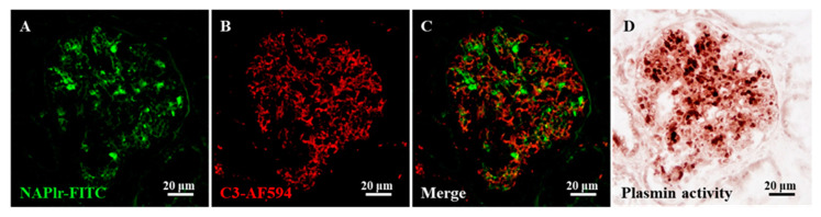
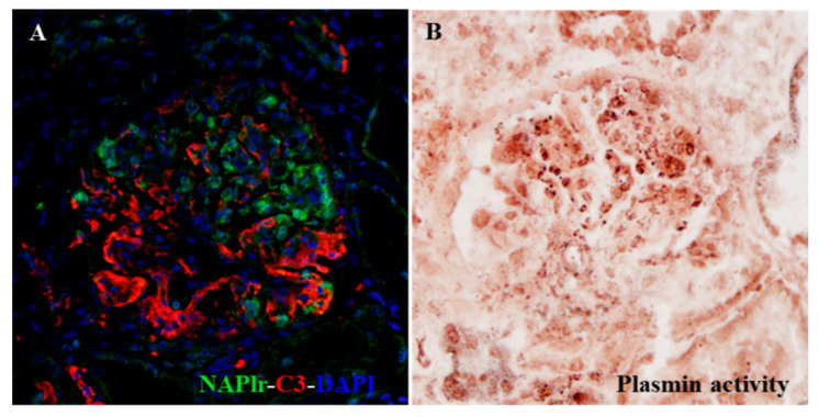
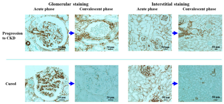
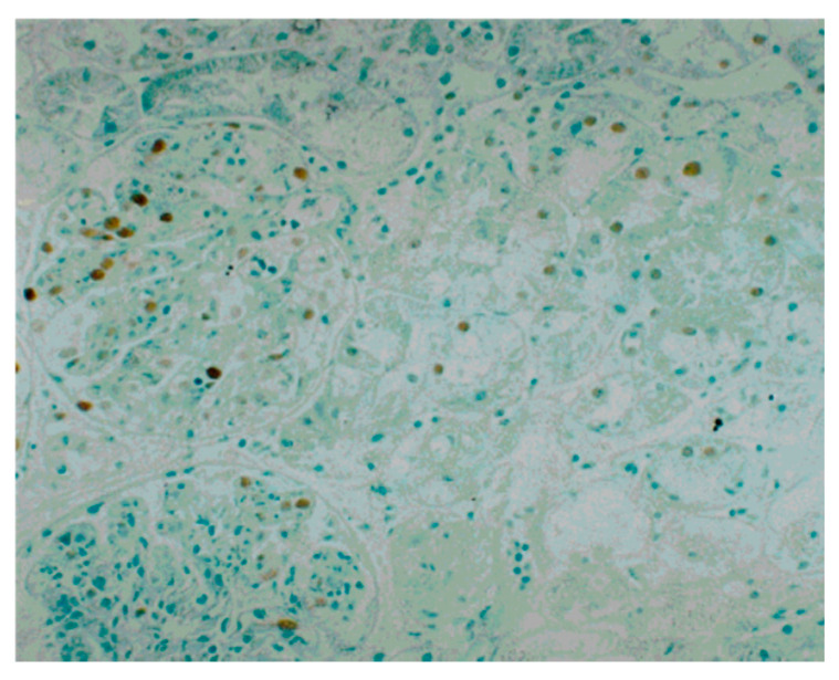
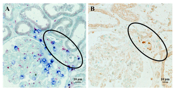
Acute glomerulonephritis (AGN) triggered by infection is still one of the major causes of acute kidney injury. During the previous two decades, there has been a major paradigm shift in the epidemiology of AGN. The incidence of poststreptococcal acute glomerulonephritis (PSAGN), which develops after the cure of group A Streptococcus infection in children has decreased, whereas adult AGN cases have been increasing, and those associated with nonstreptococcal infections, particularly infections by Staphylococcus, are now as common as PSAGN. In adult AGN patients, particularly older patients with comorbidities, infections are usually ongoing at the time when glomerulonephritis is diagnosed; thus, the term "infection-related glomerulonephritis (IRGN)" has recently been popularly used instead of "post-infectious AGN". The prognosis of children with PSAGN is generally considered excellent compared with that of adult IRGN cases. However, long-term epidemiological analysis demonstrated that an episode of PSAGN in childhood is a strong risk factor for chronic kidney disease (CKD), even after the complete remission of PSAGN. Although the precise mechanism of the transition from IRGN to CKD remains unknown, its clarification is important as it will lead to the prevention of CKD. In this review, we therefore focus on the possible factors that may contribute to the progression of IRGN into CKD. Four factors, namely, persistent infection, genetic background of the host's complement system, tubulointerstitial changes, and pre-existing histological damage, are discussed.

Keywords: acute kidney injury; alpha-smooth muscle actin; chronic kidney disease; infection-related glomerulonephritis; nephritis-associated plasmin receptor; poststreptococcal acute glomerulonephritis; tubulo-interstitial change.


Conflict of interest statement

The authors declare that they have no conflict of interest associated with this manuscript.

Figures

Figure 1
Figure 1
Summary of the concept of this review. Induction of IRGN and its outcome are summarized. IRGN may cure completely or may progress into CKD. Four factors that may contribute to the progression of IRGN into CKD are depicted. Solid arrow indicates the main flow of induction of IRGN and its outcome. While dotted arrow indicate the flow of patients with pre-existing comorbidities (Factor 4). NAPlr: nephritis-associated plasmin receptor; IRGN: infection-related glomerulonephritis; PGN: proliferative glomerulonephritis; GN: glomerulonephritis; C activation: complement activation; MPGN: membranoproliferative glomerulonephritis; MesPGN: mesangial proliferative glomerulonephritis; T-I: tubulo-interstitial; C3-G: C3 glomerulopathy; PIGN: postinfectious glomerulonephritis; CKD: chronic kidney disease.
Figure 2
Figure 2
Representative photomicrographs of the histological staining for C3, nephritis-associated plasmin receptor (NAPlr), and plasmin activity in the glomeruli of a post-streptococcal acute glomerulonephritis (PSAGN) patient. (AC) Double immunofluorescence (IF) staining for NAPlr (fluorescein isothiocyanate, green) and complement C3 (Alexa Fluor 594, red). Both NAPlr (A) and C3 (B) were positive in the glomeruli, but they generally were not colocalized, as shown in the merged image (C). (D) Plasmin activity assessed by in situ zymography on a serial section was found to be positive, and to have a similar distribution to the NAPlr staining in the glomeruli. Details of all staining methods have been described previously [11].
Figure 3
Figure 3
Photomicrographs of histological staining for C3, NAPlr, and plasmin activity in the glomeruli of a patient with infection-related glomerulonephritis (IRGN) induced by asymptomatic sinusitis [36]. Although the infection was clinically inapparent, double IF staining for NAPlr (fluorescein isothiocyanate, green) and C3 (Alexa Fluor 594, red) with nuclear staining for DAPI (blue) showed glomerular deposition of NAPlr and C3 (A). Furthermore, glomerular plasmin activity assessed by in situ zymography on a serial section demonstrated a similar distribution as NAPlr deposition, providing histological evidence for the substantial involvement of bacterial infection in the development of glomerulonephritis (B).
Figure 4
Figure 4
Immunoperoxidase staining for alpha-smooth muscle actin (α-SMA) with methyl green counterstaining in repeated renal biopsy sections from the acute phase and the convalescent phase of a PSAGN patient who developed chronic kidney disease (CKD), and a PSAGN patient who was cured. Strong positive staining of α-SMA in the mesangial area of the glomeruli was observed in tissues of PSAGN patients in the acute phase irrespective of disease prognosis, and this glomerular staining diminished uniformly in the convalescent phase irrespective of disease prognosis. Interstitial α-SMA staining was more intense in the PSAGN patient who developed CKD than in the patient who was cured, both in the acute phase and in the convalescent phase. For the indirect immunoperoxidase staining for α-SMA, a mouse monoclonal antibody (clone 1A4) was used as the primary antibody, and a peroxidase-conjugated goat anti-mouse IgG antibody was used as the secondary antibody.
Figure 5
Figure 5
Immunoperoxidase staining for proliferating cell nuclear antigen (PCNA) and counterstaining with methyl green in a renal biopsy tissue from a PSAGN patient (original magnification, ×100) [23]. Prominent expression of PCNA in the glomerulus and tubular epithelial cells was observed. The distribution of PCNA-positive cells was variable; i.e., there was a considerable difference in the number of PCNA-positive cells between each glomerulus and between different parts of the tubules.
Figure 6
Figure 6
Staining of infiltrating cells and PCNA in serial sections of renal biopsy tissue from a patient with PSAGN. Double staining for neutrophils (chloroesterase staining: blue) and macrophages (immuno-alkaline phosphatase staining for CD11c: red) demonstrated extensive glomerular infiltration of both cell types, which spread out from the vascular pole (circled region) (A). Tubular epithelial cells of the same region (circled) showed positive PCNA staining (immunoperoxidase staining: brown) on a serial section (B). Details of all the staining methods have been described previously [22].

Similar articles

Cited by

References

    1. Coca S.G., Singanamala S., Parikh C.R. Chronic kidney disease after acute kidney injury: A systematic review and meta-analysis. Kidney Int. 2012;81:442–448. doi: 10.1038/ki.2011.379. - DOI - PMC - PubMed
    1. Satoskar A.A., Parikh S.V., Nadasdy T. Epidemiology, pathogenesis, treatment and outcomes of infection-associated glomerulonephritis. Nat. Rev. Nephrol. 2020;16:32–50. doi: 10.1038/s41581-019-0178-8. - DOI - PubMed
    1. Nasr S.H., Radhakrishnan J., D’Agati V.D. Bacterial infection-related glomerulonephritis in adults. Kidney Int. 2013;83:792–803. doi: 10.1038/ki.2012.407. - DOI - PubMed
    1. Nasr S.H., Fidler M.E., Valeri A.M., Cornell L.D., Sethi S., Zoller A., Stokes M.B., Markowitz G.S., D’Agati V.D. Postinfectious glomerulonephritis in the elderly. J. Am. Soc. Nephrol. 2011;22:187–195. doi: 10.1681/ASN.2010060611. - DOI - PubMed
    1. Baldwin D.S., Gluck M.C., Schacht R.G., Gallo G. The long-term course of poststreptococcal glomerulonephritis. Ann. Intern. Med. 1974;80:342–358. doi: 10.7326/0003-4819-80-3-342. - DOI - PubMed

MeSH terms

LinkOut - more resources