Review
. 2021 Jan 18;22(2):926.
doi: 10.3390/ijms22020926.

Functions of Muscarinic Receptor Subtypes in Gastrointestinal Smooth Muscle: A Review of Studies with Receptor-Knockout Mice

Affiliations
Review

Functions of Muscarinic Receptor Subtypes in Gastrointestinal Smooth Muscle: A Review of Studies with Receptor-Knockout Mice

Yasuyuki Tanahashi et al. Int J Mol Sci. .

Abstract

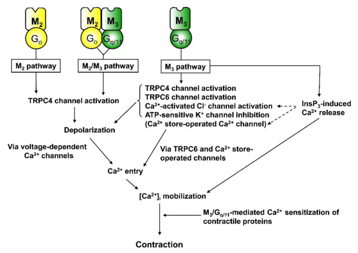
Parasympathetic signalling via muscarinic acetylcholine receptors (mAChRs) regulates gastrointestinal smooth muscle function. In most instances, the mAChR population in smooth muscle consists mainly of M2 and M3 subtypes in a roughly 80% to 20% mixture. Stimulation of these mAChRs triggers a complex array of biochemical and electrical events in the cell via associated G proteins, leading to smooth muscle contraction and facilitating gastrointestinal motility. Major signalling events induced by mAChRs include adenylyl cyclase inhibition, phosphoinositide hydrolysis, intracellular Ca2+ mobilisation, myofilament Ca2+ sensitisation, generation of non-selective cationic and chloride currents, K+ current modulation, inhibition or potentiation of voltage-dependent Ca2+ currents and membrane depolarisation. A lack of ligands with a high degree of receptor subtype selectivity and the frequent contribution of multiple receptor subtypes to responses in the same cell type have hampered studies on the signal transduction mechanisms and functions of individual mAChR subtypes. Therefore, novel strategies such as genetic manipulation are required to elucidate both the contributions of specific AChR subtypes to smooth muscle function and the underlying molecular mechanisms. In this article, we review recent studies on muscarinic function in gastrointestinal smooth muscle using mAChR subtype-knockout mice.

Keywords: gastrointestinal tract; knockout mouse; muscarinic receptor subtypes; non-selective cationic channels; signal transduction pathways; smooth muscle.


Conflict of interest statement

No conflict of interest, financial or otherwise, are declared by the authors.

Figures

Figure 1
Figure 1
Three distinct muscarinic signalling pathways leading to transient receptor potential (TRP)-like cationic channel opening in ileal myocytes (redrawn from Tanahashi et al. 2020 [39]). The M3/Gq/11/phospholipase C (PLC) pathway activates brief opening states of 70-pS and 120-pS cationic channels and concurrently evokes InsP3-induced Ca2+ release. Opening of the lower conductance channel is induced by relief from PIP2 inhibition following PLC-mediated hydrolysis, while the higher conductance channel is activated by PLC-generated diacylglycerol (DAG). The M2 pathway transmits M2 signals via Gi/o proteins to the 70-pS channel, which shifts the gating state from the brief to a longer opening mode, and also inhibits adenylyl cyclase. The M2/M3 pathway transmits M2 signals via Go protein, and M3 signals via Gq/11/PLC, to the 70-pS cationic channel, resulting in channel gating with a much longer open mode. This pathway is the major contributor to the generation of mIcat, but is inactive when either the M2 or M3 receptor is absent, or when either Go, Gq/11 or PLC is inactivated. In other words, the activity of this pathway is conditional, occurring only when both M2/Go and M3/Gq/11 signalling pathways are activated. Studies of mAChR-KO mice [23,44] and TRPC-mutant mice [42] indicate that these 70-pS and 120-pS cationic channel activities are mediated by TRPC4 and TRPC6, respectively. The M2/M3 pathway, but not the M2 or M3 pathway, involves a signalling step in which Ca2+ has a potentiating effect on TRPC channel activation, suggesting that the M3 pathway may facilitate M2/M3 pathway function through InsP3-induced Ca2+ release. Whether the M2/M3 pathway has a significant role in stimulating InsP3/DAG formation or inhibiting cAMP accumulation is currently unclear. One study suggested that Go protein is not involved in adenylyl cyclase inhibition by M2 receptors of intestinal smooth muscle [24]. These three pathways may also converge on voltage-dependent Ca2+ channels (VDCCs) to suppress Ca2+ influx via the same G-protein pathways mediating cationic channel activation (see Figure 10B in Tanahashi et al., 2009 [49]).
Figure 2
Figure 2
Signal transduction mechanisms underlying muscarinic contraction of mouse intestinal smooth muscle. The M3 pathway activates multiple intracellular Ca2+ mobilisation events, including Ca2+ influx via VDCCs and voltage-independent Ca2+-permeable channels and intracellular Ca2+ release. Voltage-dependent Ca2+ influx is initiated by depolarisation from opening of TRPC4 and TRPC6 cationic channels, opening of Ca2+-activated Cl channels and inhibition of KATP channels, while voltage-independent Ca2+ entry is mediated by opening of TRPC6 cationic channels and Ca2+ store-operated Ca2+ channels. In addition to Ca2+ mobilisation, Ca2+ sensitisation of contractile proteins is elicited, thereby increasing the efficiency of contraction–[Ca2+]i coupling. The M2 or M2/M3 pathways induce contraction through a simple Ca2+ mobilisation mechanism in which Ca2+ entry via VDCCs is activated by TRPC4 channel-induced depolarisation. These pathways play a major role in mediating muscarinic contraction, with the M2/M3 pathway making a relatively greater contribution at low agonist concentrations. The M2 pathway also induces contraction indirectly by inhibiting cAMP-dependent relaxation in response to adenylyl cyclase-activating agonists.
Figure 3
Figure 3
Effects of nicardipine on carbachol-evoked contractions in a wild-type mouse ileal longitudinal muscle strip. Carbachol was applied for 3 min at the indicated concentrations before (A) and after nicardipine treatment to block VDCCs (B). In (C), the recording traces in A and B are superimposed. Note that when muscarinic stimulation is weak, contraction is relatively more dependent on voltage-dependent Ca2+ entry and is more sensitive to PTX. This finding indicates that the M2/M3 (and M2) pathway has a major role in mediating the contractile response to weak muscarinic stimulation. When receptor stimulation is stronger, the M3 pathway predominates by activating multiple mechanisms for Ca2+ mobilisation as well as myofilament Ca2+ sensitisation.

Similar articles

Cited by

References

    1. Caulfield M.P. Muscarinic receptors-characterization, coupling and function. Pharmacol. Ther. 1993;58:319–379. doi: 10.1016/0163-7258(93)90027-B. - DOI - PubMed
    1. Caulfield M., Birdsall N. International Union of Pharmacology. XVII. Classification of muscarinic acetylcholine receptors. Pharmacol. Rev. 1998;50:279–290. - PubMed
    1. Felder C.C. Muscarinic acetylcholine-receptors-signal-transduction through multiple effectors. FASEB J. 1995;9:619–625. doi: 10.1096/fasebj.9.8.7768353. - DOI - PubMed
    1. Wess J. Molecular biology of muscarinic acetylcholine receptors. Crit. Rev. Neurobiol. 1996;10:69–99. doi: 10.1615/CritRevNeurobiol.v10.i1.40. - DOI - PubMed
    1. Jositsch G., Papadakis T., Haberberger R., Wolff M., Wess J., Kummer W. Suitability of muscarinic acetylcholine receptor antibodies for immunohistochemistry evaluated on tissue sections of receptor gene-deficient mice. Naunyn Schmiedebergs Arch. Pharmacol. 2009;379:389–395. doi: 10.1007/s00210-008-0365-9. - DOI - PMC - PubMed

MeSH terms

LinkOut - more resources