. 2021 Jan 19;22(2):942.
doi: 10.3390/ijms22020942.

Extracellular Prolidase (PEPD) Induces Anabolic Processes through EGFR, β1-integrin, and IGF-1R Signaling Pathways in an Experimental Model of Wounded Fibroblasts

Affiliations

Extracellular Prolidase (PEPD) Induces Anabolic Processes through EGFR, β1-integrin, and IGF-1R Signaling Pathways in an Experimental Model of Wounded Fibroblasts

Weronika Baszanowska et al. Int J Mol Sci. .

Abstract

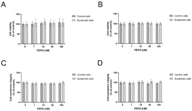
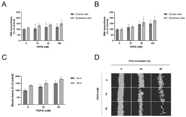
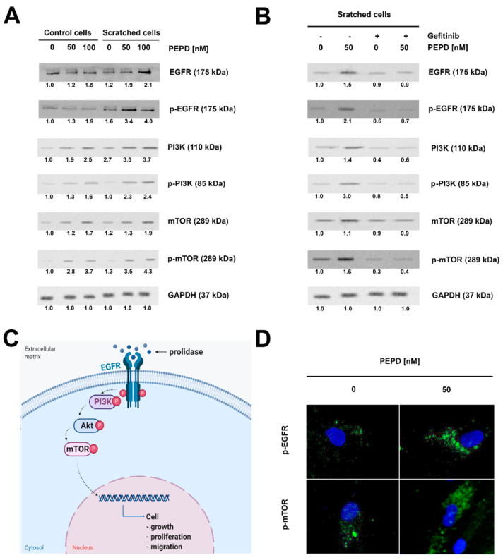
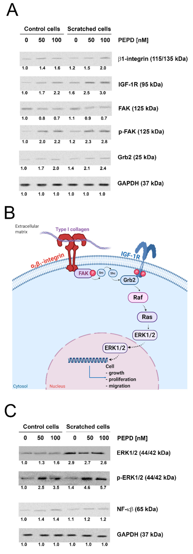
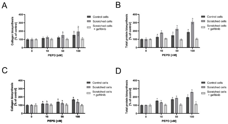
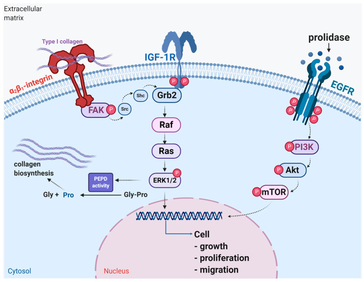
The role of prolidase (PEPD) as a ligand of the epidermal growth factor receptor (EGFR) was studied in an experimental model of wound healing in cultured fibroblasts. The cells were treated with PEPD (1-100 nM) and analysis of cell viability, proliferation, migration, collagen biosynthesis, PEPD activity, and the expressions of EGFR, insulin-like growth factor 1 (IGF-1), and β1-integrin receptor including downstream signaling proteins were performed. It has been found that PEPD stimulated proliferation and migration of fibroblasts via activation of the EGFR-downstream PI3K/Akt/mTOR signaling pathway. Simultaneously, PEPD stimulated the expression of β1-integrin and IGF-1 receptors and proteins downstream to these receptors such as FAK, Grb2, and ERK1/2. Collagen biosynthesis was increased in control and "wounded" fibroblasts under PEPD treatment. The data suggest that PEPD-induced EGFR signaling may serve as a new attempt to therapy wound healing.

Keywords: EGFR; IGF-1; PEPD; fibroblasts; prolidase; wound healing; β1-integrin.


Conflict of interest statement

The authors declare no conflict of interest.

Figures

Figure 1
Figure 1
The effect of PEPD on fibroblasts viability and cell membrane integrity. Control cells, as well as scratched fibroblasts, were treated with PEPD (1–100 nM) for 24 h (A,C) and 48 h (B,D) followed by measurement of MTT and cell membrane integrity, respectively. Mean values ± SD of three experiments done in replicates are presented. The results are significant at a, b < 0.05 indicates a vs. control (0 nM of PEPD) of control cells, b vs. control (0 nM of PEPD) of scratched cells, respectively. PEPD—prolidase.
Figure 2
Figure 2
Extracellular PEPD-dependent proliferation and migration of fibroblasts in a model of closure/scratch assay. (A,B) Control, as well as “scratched” fibroblasts, were treated with PEPD (1–100 nM) for 24 h and 48 h, and proliferation was evaluated using CyQuant Proliferation assay. (C,D) PEPD-stimulated fibroblasts migration was calculated using ImageJ software (https://imagej.nih.gov/ij/) vs. control. PEPD-treated cells were scratched and monitored using an inverted microscope (40× magnification) at 0, 24, and 48 h. Mean values ± SD of three experiments done in replicates are presented. The results are significant at a, b < 0.05, and indicates a vs. control (0 nM of PEPD) of control cells and part C of 24 h incubation, b vs. control (0 nM of PEPD) of scratched cells, and part C of 48 h incubation, respectively. PEPD—prolidase.
Figure 3
Figure 3
Extracellular PEPD-induced epidermal growth factor receptor (EGFR)-downstream signaling pathway. (A) Western blot for the proteins of EGFR-downstream signaling pathway in lysates of control and “scratched” PEPD-treated fibroblasts (PEPD, 1−100 nM) for 24 h or PEPD-treated fibroblasts (PEPD, 0 and 50 nM) with an inhibitor of EGFR (Gefitinib pretreated cells for 2 h, 0 and 45 µM) for 24 h. GAPDH was used as a loading control. (B) Representative blot images were shown (densitometry of protein stains is presented under protein bands as a ratio versus control; Supplementary Figure S1). GAPDH was used as a loading control. (C) Illustration of the PEPD-dependent EGFR-downstream signaling pathway. Created with BioRender.com. (D) Representative results of immunostaining of p-EGFR and p-mTOR in PEPD-stimulated fibroblasts (50 nM) for 24 h are presented; magnification 200×.
Figure 4
Figure 4
Extracellular PEPD induced expression of the β1-integrin receptor and IGF-1R signaling proteins in control and “scratched” fibroblast models. (A) The proteins of β1-integrin receptor and IGF-1R downstream signaling pathways, FAK, Grb2, and (C) NF-ĸβ and ERK1/2 were analyzed by Western blot in lysates of PEPD-treated fibroblasts (50, and 100 nM). Representative blot images were shown (densitometry of protein stains is presented under protein bands as a ratio versus control; Supplementary Figure S2). GAPDH was used as a loading control. (B) Illustration of the β1-integrin receptor-downstream signaling pathway. Created with BioRender.com.
Figure 5
Figure 5
Extracellular PEPD activated collagen and total protein biosynthesis in control and “scratched” fibroblast models. Collagen biosynthesis (A,C) and total protein biosynthesis (B,D) in PEPD-treated fibroblasts (1–100 nM) in the presence and absence of EGFR inhibitor (Gefitinib, 0 and 45 µM pretreated cells for 2 h) after 24 and 48 h incubation, respectively. The values were presented as a percent of control cells (0 nM of PEPD). Mean values± SD of three experiments done in replicates is presented. The results are significant at a, b, c < 0.05, and are marked as a vs. control (0 nM of PEPD) of control cells, b vs. control (0 nM of PEPD) of scratched cells, c vs. control (0 nM of PEPD) of scratched cells incubated with gefitinib, respectively. PEPD—prolidase.
Figure 6
Figure 6
Schematic representation of the functional significance of activation of EGFR, IGF-1R, and β1-integrin receptor downstream signaling in wound healing. Under experimental conditions of mechanically “wounded” fibroblasts, PEPD activates EGFR-dependent downstream PI3K/Akt/mTOR signaling, while IGF-1R and β1-integrin receptor cooperate to activate MAPK (ERK1/2) pathway resulting in increased cell proliferation, migration and collagen biosynthesis. Created with BioRender.com.

References

    1. Leavitt T., Hu M.S., Marshall C.D., Barnes L.A., Lorenz H.P., Longaker M.T. Scarless wound healing: Finding the right cells and signals. Cell Tissue Res. 2016;365:483–493. doi: 10.1007/s00441-016-2424-8. - DOI - PMC - PubMed
    1. Gurtner G.C., Werner S., Barrandon Y., Longaker M.T. Wound repair and regeneration. Nature. 2008;453:314–321. doi: 10.1038/nature07039. - DOI - PubMed
    1. Rose L.F., Chan R.K. The Burn Wound Microenvironment. Adv. Wound Care. 2016;5:106–118. doi: 10.1089/wound.2014.0536. - DOI - PMC - PubMed
    1. Palka J., Adelmann-Grill B.C., Francz P.I., Bayreuther K. Differentiation stage and cell cycle position determine the chemotactic response of fibroblasts. Folia Histochem. Cytobiol. 1996;34:121–127. - PubMed
    1. Pierard G.E., Cornil F., Lapiere C.M. Pathogenesis of ulcerations in deficiency of prolidase. The role of angiopathy and of deposits of amyloid. Am. J. Dermatopathol. 1984;6:491–497. doi: 10.1097/00000372-198410000-00013. - DOI - PubMed

LinkOut - more resources