. 2021 Jan 19;11(1):124.
doi: 10.3390/biom11010124.

Modeling the Influenza A NP-vRNA-Polymerase Complex in Atomic Detail

Affiliations

Modeling the Influenza A NP-vRNA-Polymerase Complex in Atomic Detail

Jacob C Miner et al. Biomolecules. .

Abstract

Seasonal flu is an acute respiratory disease that exacts a massive toll on human populations, healthcare systems and economies. The disease is caused by an enveloped Influenza virus containing eight ribonucleoprotein (RNP) complexes. Each RNP incorporates multiple copies of nucleoprotein (NP), a fragment of the viral genome (vRNA), and a viral RNA-dependent RNA polymerase (POL), and is responsible for packaging the viral genome and performing critical functions including replication and transcription. A complete model of an Influenza RNP in atomic detail can elucidate the structural basis for viral genome functions, and identify potential targets for viral therapeutics. In this work we construct a model of a complete Influenza A RNP complex in atomic detail using multiple sources of structural and sequence information and a series of homology-modeling techniques, including a motif-matching fragment assembly method. Our final model provides a rationale for experimentally-observed changes to viral polymerase activity in numerous mutational assays. Further, our model reveals specific interactions between the three primary structural components of the RNP, including potential targets for blocking POL-binding to the NP-vRNA complex. The methods developed in this work open the possibility of elucidating other functionally-relevant atomic-scale interactions in additional RNP structures and other biomolecular complexes.

Keywords: Influenza A; RNP; homology modeling; viral genome.


Conflict of interest statement

The authors declare no conflict of interest.

Figures

Figure 1
Figure 1
Single-stranded RNA structure prediction. (a) The α and β torsional angles for each nucleotide in T. thermophilus 30S rRNA shows a linear correlation, allowing an initial prediction of nucleobase orientation based on the backbone torsion. (b) Four target phosphorous atoms (in yellow) of vRNA at the NP surface align to the phosphorous atoms of a template structure ((c), in magenta), from which the embedded nucleoside structure is derived. (d) By repeating this method across the NP surface a model of the vRNA is generated.
Figure 2
Figure 2
NP dimer arrangements. (a) Two interacting NP molecules from the trimer conformation (Protein Data Bank (PDB) accession code: 2IQH) are shown with the folded domain (residues 402-421) from one nucleoprotein (NP) (cyan) occupying the binding pocket of the second NP (yellow). Dotted lines between the folded domain and the main NP body are shown as a guide to the eye. (b) All three NP dimer arrangements are shown: trimer (cyan), circular (magenta), and helical (green) from PDB accession codes 2IQH, 2WFS, and 4BBL, respectively. The receiving NP (yellow), where the folded domain enters the binding pocket are superimposable in all three arrangements (red). (c) An enlarged view of the folded domain and the orientations of the trimer (cyan), circular (magenta), and helical (green) NPs shows the expected connectivity.
Figure 3
Figure 3
RNP complex. (a) The hairpin model of RNP is composed of two arrangements of NP oligomers (b): circular (gray) and helical (brown, yellow) suprastructures. (c) The two suprastructures (PDB accession codes: 2WFS, 4BBL) are mapped onto one another using two NPs in each structure (circular: A& I, helical: W& X). (d) After overlaying these NPs, one NP from each suprastructure is retained in the final model. (e) The close alignment of NP-X and NP-I means that either can be selected for the full RNP model. However, due to the orientations of the folded domain from NP-U and the binding pocket of NP-B, only NP-A can form a link between both NPs and unite the two suprastructures.
Figure 4
Figure 4
Bridging vRNA between two NP molecules. A five-residue loop (residues 230–234 from T. thermophilus 30S ribosomal subunit, PDB accession code: 1J5E) aligns with the vRNA nucleotides at the junction of the helical and circular suprastructures. The aligned residues of this RNA loop and the NP-bound vRNA (344 and 348) are shown in green, bridging residues are shown in magenta, and NP-associated vRNA residues are shown in cyan. The NP molecules are shown in yellow.
Figure 5
Figure 5
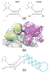
Modeling POL within the RNP complex. (a) The “half-corkscrew” secondary structure of vRNA (PDB accession code: 4WSB) is used to model POL-binding at the 3 and 5 termini. (b) Docking POL into the cryo-EM density map shows the vRNA promoter structure (blue) occupies the space between the NP—vRNA complex (yellow and cyan, respectively) and POL (magenta). A test of the goodness-of-fit for POL identifies 78.4% of the heavy atoms are retained within the outer contour of the EM density map of emd_2208 [16]. (c) Both ends of the vRNA in the “half-corkscrew” arrangement are joined with the rest of the vRNA at the residues shown.
Figure 6
Figure 6
Structural model of the Influenza A NP–vRNA–polymerase (POL) complex. This model corresponds to the NS1 segment of the Influenza A/Albany/3/1967 RNP. Different NP oligomeric suprastructures are plotted as follows: helical NPs 1–12 (shown as yellow ribbons), circular NPs 13–21 (shown as gray ribbons), and helical NPs 22–33 (shown as tan calotte/space-filling). The vRNA (cyan) is plotted along the length of the RNP, and POL is shown in magenta.
Figure 7
Figure 7
Interactions between POL and NP–vRNA. (a) The folded domain from NP-33 (residues 405–422, yellow) interacts with PB1 (cyan) and PB2 (magenta) of the polymerase. Residue R416 (blue) of NP-33, and E18 (red) of PB2 form a salt bridge. (b) Additional salt bridges are formed by the 3 end of vRNA (cyan) to the polymerase subunits: A830 to K209 of PA (shown in yellow), and U853 to R512 within a hairpin structure of PB1 (shown in tan). (c) K198 of PB1 (yellow) points away from vRNA, while the K198R mutation (magenta) allows for a salt bridge to form with the phosphate group of C842 of the vRNA. The larger R198 side-chain thus causes an increase in POL-binding to vRNA.

References

    1. Bedford T., Riley S., Barr I.G., Broor S., Chadha M., Cox N.J., Daniels R.S., Gunasekaran C.P., Hurt A.C., Kelso A., et al. Global circulation patterns of seasonal influenza viruses vary with antigenic drift. Nature. 2015;523:217–220. doi: 10.1038/nature14460. - DOI - PMC - PubMed
    1. Iuliano A.D., Roguski K.M., Chang H.H., Muscatello D.J., Palekar R., Tempia S., Cohen C., Gran J.M., Schanzer D., Cowling B.J., et al. Estimates of global seasonal influenza-associated respiratory mortality: A modelling study. Lancet. 2018;391:1285–1300. doi: 10.1016/S0140-6736(17)33293-2. - DOI - PMC - PubMed
    1. Pflug A., Lukarska M., Resa-Infante P., Reich S., Cusack S. Structural insights into RNA synthesis by the influenza virus transcription-replication machine. Virus Res. 2017;234:103–117. doi: 10.1016/j.virusres.2017.01.013. - DOI - PubMed
    1. Chaimayo C., Hayashi T., Underwood A., Hodges E., Takimoto T. Selective incorporation of vRNP into influenza A virions determined by its specific interaction with M1 protein. Virology. 2017;505:23–32. doi: 10.1016/j.virol.2017.02.008. - DOI - PMC - PubMed
    1. Zheng W., Tao Y.J. Structure and assembly of the influenza A virus ribonucleoprotein complex. FEBS Lett. 2013;587:1206–1214. doi: 10.1016/j.febslet.2013.02.048. - DOI - PubMed

Publication types

LinkOut - more resources