. 2021 Jan 19;22(2):955.
doi: 10.3390/ijms22020955.

Mechanical Stress Induces Ca2+-Dependent Signal Transduction in Erythroblasts and Modulates Erythropoiesis

Affiliations

Mechanical Stress Induces Ca2+-Dependent Signal Transduction in Erythroblasts and Modulates Erythropoiesis

Francesca Aglialoro et al. Int J Mol Sci. .

Abstract

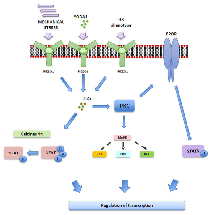
Bioreactors are increasingly implemented for large scale cultures of various mammalian cells, which requires optimization of culture conditions. Such upscaling is also required to produce red blood cells (RBC) for transfusion and therapy purposes. However, the physiological suitability of RBC cultures to be transferred to stirred bioreactors is not well understood. PIEZO1 is the most abundantly expressed known mechanosensor on erythroid cells. It is a cation channel that translates mechanical forces directly into a physiological response. We investigated signaling cascades downstream of PIEZO1 activated upon transitioning stationary cultures to orbital shaking associated with mechanical stress, and compared the results to direct activation of PIEZO1 by the chemical agonist Yoda1. Erythroblasts subjected to orbital shaking displayed decreased proliferation, comparable to incubation in the presence of a low dose of Yoda1. Epo (Erythropoietin)-dependent STAT5 phosphorylation, and Calcineurin-dependent NFAT dephosphorylation was enhanced. Phosphorylation of ERK was also induced by both orbital shaking and Yoda1 treatment. Activation of these pathways was inhibited by intracellular Ca2+ chelation (BAPTA-AM) in the orbital shaker. Our results suggest that PIEZO1 is functional and could be activated by the mechanical forces in a bioreactor setup, and results in the induction of Ca2+-dependent signaling cascades regulating various aspects of erythropoiesis. With this study, we showed that Yoda1 treatment and mechanical stress induced via orbital shaking results in comparable activation of some Ca2+-dependent pathways, exhibiting that there are direct physiological outcomes of mechanical stress on erythroblasts.

Keywords: PIEZO1; calcium signal transduction; mechanical stress.


Conflict of interest statement

The authors declare no relevant conflict of interest.

Figures

Figure 1
Figure 1
Mechanically stimulated and Yoda1-treated erythroblasts proliferation is impaired. Erythroblasts were cultured in flasks on the orbital shaker at 300 RPM (Flask: F) and in dishes in static control condition (Dish Control: C) for 4 days (A,C,E). In parallel, erythroblasts were cultured in absence (Control: C) or the presence of 0.2 μM (0.2), 1 μM (1) and 5 μM (5) Yoda1 or the solvent DMSO (DMSO: D) (A,B,D,F) Cell counts were assessed daily, cell number was maintained below 1,5 × 10⁶ by daily dilution and cumulative cells were calculated (depicted as fold change (FC)). (C,D) Cell death was assessed by Propidium Iodide (PI) staining at day 0, 2, or 4 for orbital shaking, and at day 0 and 4 for Yoda1 treatment. Significance refers to the most mature CD235a+/CD49d- population. (E,F) Erythroblasts were stained with Pacific Blue-labeled anti CD49d and PE-labeled anti CD235a (GPA) antibodies and analyzed on the flow cytometer. Expression is plotted as percentage of more immature (black; CD235a-/CD49d+), and more mature erythroblasts (light grey; CD235a+/CD49d+ and dark grey; CD235a+/CD49d-). All experiments were performed in triplicates, error bars indicate standard deviation. For orbital shaker experiments paired t-tests, and for Yoda1 experiments unpaired t-tests were performed to assay significance between conditions (* p < 0.05; ** p < 0.01; *** p < 0.001; **** p < 0.0001).
Figure 2
Figure 2
Mechanical stimulation and Yoda1 incubation induces Ca2+-dependent signaling pathways. Erythroblasts were either cultured on an orbital shaker set at 300 RPM and sampled at 0, 10, 20, 40, and 60 min, or stimulated with 1 μM Yoda1 with or without specific inhibitors and sampled at 0, 10, 30, 60 min. Total protein lysates were subjected to SDS-polyacrylamide gel electrophoresis and western blotting. (A) Some of the erythroblasts were pretreated with 1 μM FK506 before Yoda1 incubation. Blots were stained with antibodies against NFATc2 (arrows indicate the slower migrating phosphorylated, and faster migrating unphosphorylated isoforms). (B) Some of the erythroblasts were pretreated with 500 nM Gö6976 before Yoda1 incubation. Blots were stained with antibodies against tyrosine-phosphorylated-STAT5 (pSTAT5) and total STAT5 (t-STAT5). (C) Blots were stained with antibodies against phosphorylated JNK (Thr183/Tyr185) and total JNK, phosphorylated ERK1/2 (Thr202/Tyr204) and total ERK1/2, phosphorylated P38 (Thr180/Tyr182) and total p38. With orbital shaking even after a long exposure, nothing other than background bands were visible for phosphorylated JNK
Figure 3
Figure 3
Intracellular Ca2+ chelation prevents shear-induced signaling pathways. Cultured erythroblasts were preincubated for 10 min under static conditions, and subsequently orbitally shaken at 300 rpm for 20 or 60 min (first lane = Time 0). These cultures were left untreated (CTRL, -), treated with solvent DMSO (CTRL, D), or pre-incubated with 1, 3, and 10 μM of the Ca2+ chelator BAPTA-AM (BAPTA(μM), 1, 3, 10). Total protein lysates were subjected to SDS-polyacrylamide gel electrophoresis and western blotting. (A) Blots were stained with anti-NFATc2, anti-p-STAT5 and total-STAT5, anti-p-ERK1/2 (Thr202/Tyr204), anti-Total-ERK1/2, anti-p-P38 (Thr180/Tyr182), anti-total p38. (B) Blots were stained with anti-pJNK (Thr183/Tyr185) and anti-Total-JNK.
Figure 4
Figure 4
Overview of possible signal transduction downstream of PIEZO1 activation triggered by mechanical stress, upon the PIEZO1 agonist Yoda1 or the HX phenotype.

References

    1. Zeuner A., Martelli F., Vaglio S., Federici G.F., Whitsett C., Migliaccio A.R. Concise Review: Stem Cell-Derived Erythrocytes as Upcoming Players in Blood Transfusion. Stem Cells. 2012;30:1587–1596. doi: 10.1002/stem.1136. - DOI - PMC - PubMed
    1. Pishesha N., Bilate A.M., Wibowo M.C., Huang N.J., Li Z., Deshycka R., Bousbaine D., Li H. Engineered erythrocytes covalently linked to antigenic peptides can protect against autoimmune dis-ease. Proc. Natl. Acad. Sci. USA. 2017;114:3157–3162. doi: 10.1073/pnas.1701746114. - DOI - PMC - PubMed
    1. Wang C., Ye Y., Sun W., Yu J., Wang J., Lawrence D.S., Buse J.B., Gu Z. Red Blood Cells for Glucose-Responsive Insulin Delivery. Adv. Mater. 2017;29 doi: 10.1002/adma.201606617. - DOI - PubMed
    1. Huang N.J., Pishesha N., Mukherjee J., Zhang S., Deshycka R., Sudaryo V., Dong M., Shoemaker C.B., Lodish H.F. Genetically engineered red cells expressing single domain camelid antibodies confer long-term protec-tion against botulinum neurotoxin. Nat. Commun. 2017;8:423. doi: 10.1038/s41467-017-00448-0. - DOI - PMC - PubMed
    1. Giarratana M.-C., Marie T., Darghouth D., Douay L. Biological validation of bio-engineered red blood cell productions. Blood Cells Mol. Dis. 2013;50:69–79. doi: 10.1016/j.bcmd.2012.09.003. - DOI - PubMed

MeSH terms

LinkOut - more resources