Review
. 2021 Jan 19;22(2):963.
doi: 10.3390/ijms22020963.

Exosomes: A Key Piece in Asthmatic Inflammation

Affiliations
Review

Exosomes: A Key Piece in Asthmatic Inflammation

José A Cañas et al. Int J Mol Sci. .

Abstract

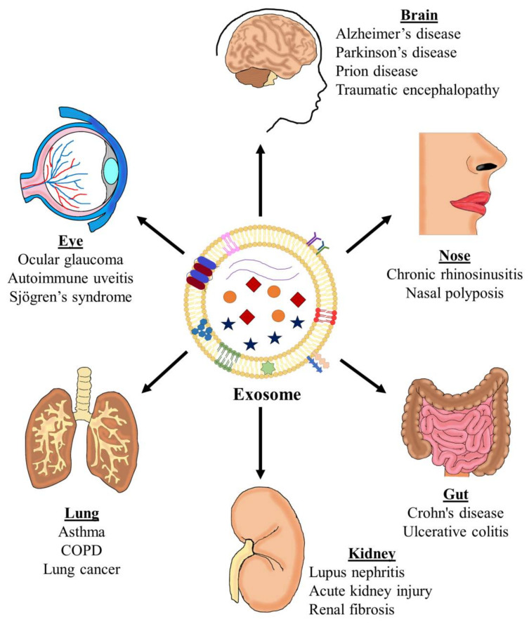
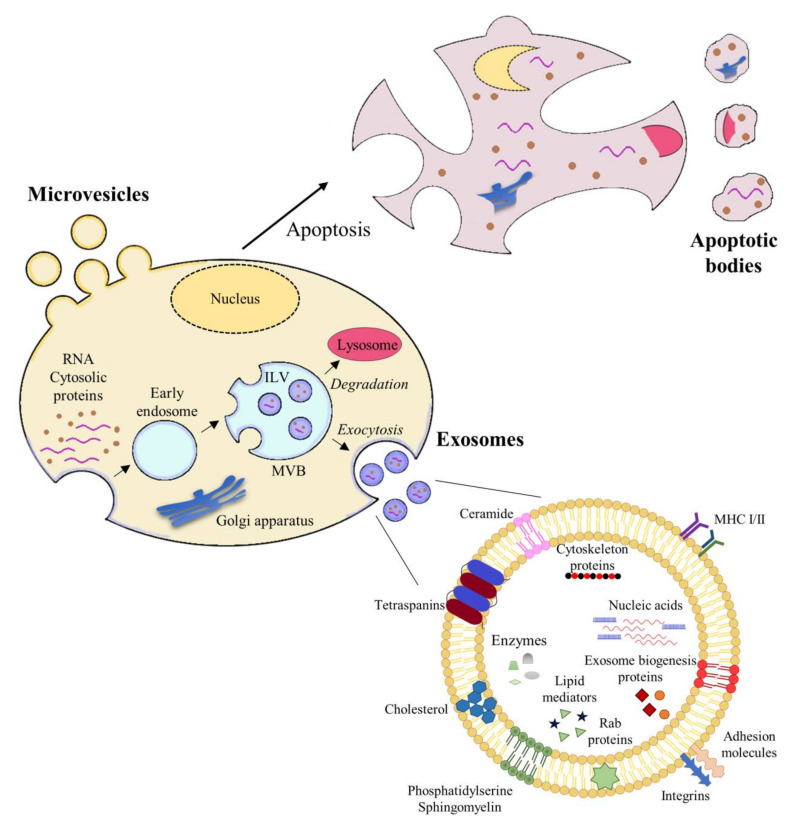
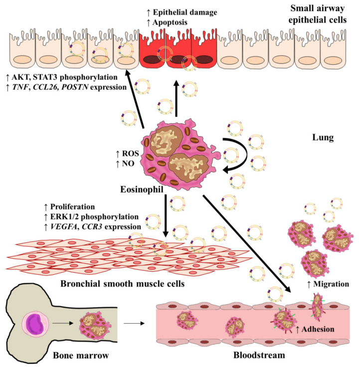
Asthma is a chronic disease of the airways that has an important inflammatory component. Multiple cells are implicated in asthma pathogenesis (lymphocytes, eosinophils, mast cells, basophils, neutrophils), releasing a wide variety of cytokines. These cells can exert their inflammatory functions throughout extracellular vesicles (EVs), which are small vesicles released by donor cells into the extracellular microenvironment that can be taken up by recipient cells. Depending on their size, EVs can be classified as microvesicles, exosomes, or apoptotic bodies. EVs are heterogeneous spherical structures secreted by almost all cell types. One of their main functions is to act as transporters of a wide range of molecules, such as proteins, lipids, and microRNAs (miRNAs), which are single-stranded RNAs of approximately 22 nucleotides in length. Therefore, exosomes could influence several physiological and pathological processes, including those involved in asthma. They can be detected in multiple cell types and biofluids, providing a wealth of information about the processes that take account in a pathological scenario. This review thus summarizes the most recent insights concerning the role of exosomes from different sources (several cell populations and biofluids) in one of the most prevalent respiratory diseases, asthma.

Keywords: asthma; biofluids; eosinophils; exosomes; extracellular vesicles; miRNAs.


Conflict of interest statement

V.d.P. has received honoraria (advisory board, speaker) and/or institutional grant/research support from Astra-Zeneca and GSK. The rest of the authors declare no conflicts of interest.

Figures

Figure 1
Figure 1
The implication of exosomes in inflammatory diseases. Exosomes have been described as modulators of different inflammatory diseases in different organs, including the brain (Alzheimer’s and Parkinson’s diseases), eye (ocular glaucoma), nose (chronic rhinosinusitis, nasal polyposis), lung (asthma, chronic obstructive pulmonary disease (COPD), and lung cancer), gut (inflammatory bowel disease), and kidney (lupus nephritis, acute kidney injury, and renal fibrosis). Figures inside and in the surface of the exosome represent some of their components. Inside: blue star: lipid mediators; exosome biogenesis proteins; violet lines: nucleic acids. In the surface: blue and red elipses: tetraspanins; green, red and pink structures: ceramide, phosphatidylserine, and sphingomyelin; violet and green: major histocompatibility complex (MHC)-I and -II, blue structure: integrin; pink structure; adhesion molecule; blue hexagons: cholesterol; green star: Rab proteins.
Figure 2
Figure 2
Extracellular vesicle biogenesis and exosome structure. Cells release several types of extracellular vesicles (EVs), including apoptotic bodies, microvesicles, and exosomes. Apoptotic bodies are large vesicles (1000–5000 nm in diameter), which are released from apoptotic cells. Microvesicles are medium-large EVs (100 nm to 1 µm in diameter), which originate directly from the cell membrane, followed by fission and release towards the extracellular space. Exosomes are small vesicles (30–150 nm) derived from endosomes. Maturation of early endosomes into late endosomes produces invaginations called intraluminal vesicles (ILVs). This endosome with ILVs is denominated multivesicular bodies (MVBs). These MVBs are able to fuse with lysosomes and degrade their cargo or with the plasma membrane to release ILVs towards the extracellular space. Exosomes are formed by a double lipid membrane and contain cytoskeletal proteins, tetraspanins, integrins, and adhesion molecules, and other proteins that reflect their endosomal biogenesis.
Figure 3
Figure 3
Specific roles of eosinophil-derived exosomes in asthmatic inflammation. Exosomes released by eosinophils are able to alter several functions associated with asthmatic pathology on both eosinophils themselves and structural lung cells, such as small airway epithelial cells and smooth bronchial muscle cells. Exosomes can increase eosinophil adhesion and migration and the release of ROS and NO, contributing to tissue damage. Moreover, exosomes augment BSMC proliferation and SAEC injury, increase the expression of several genes implicated in asthmatic inflammation and remodeling (TNF, POSTN, CCL26, POSTN, VEGFA, and CCR3), and alter some pathways implicated in asthma, including MAPK and JAK/STAT. Abbreviations. AKT: protein kinase B; STAT3: signal transducer and activator of transcription 3; TNF: tumor necrosis factor; CCL26: C-C chemokine ligand 26; POSTN: periostin; ROS: reactive oxygen species; NO: nitric oxide; ERK: extracellular regulated kinase; VEGFA: vascular endothelial growth factor A; CCR3: C-C chemokine receptor 3.

Similar articles

Cited by

References

    1. Lambrecht B.N., Hammad H. The immunology of asthma. Nat. Immunol. 2015;16:45–56. doi: 10.1038/ni.3049. - DOI - PubMed
    1. Mauer Y., Taliercio R.M. Managing adult asthma: The 2019 GINA guidelines. Clevel. Clin. J. Med. 2020;87:569–575. doi: 10.3949/ccjm.87a.19136. - DOI - PubMed
    1. Wenzel S.E. Complex phenotypes in asthma: Current definitions. Pulm. Pharmacol. Ther. 2013;26:710–715. doi: 10.1016/j.pupt.2013.07.003. - DOI - PubMed
    1. Mortaz E., Alipoor S.D., Varahram M., Jamaati H., Garssen J., Mumby S.E., Adcock I.M. Exosomes in severe asthma: Update in their roles and potential in therapy. Biomed. Res. Int. 2018;2018:2862187. doi: 10.1155/2018/2862187. - DOI - PMC - PubMed
    1. Schorey J.S., Cheng Y., Singh P.P., Smith V.L. Exosomes and other extracellular vesicles in host–pathogen interactions. EMBO Rep. 2015;16:24–43. doi: 10.15252/embr.201439363. - DOI - PMC - PubMed