Review
. 2021 Jan 19;26(2):514.
doi: 10.3390/molecules26020514.

Effects of Ionic Liquids on Metalloproteins

Affiliations
Review

Effects of Ionic Liquids on Metalloproteins

Aashka Y Patel et al. Molecules. .

Abstract

In the past decade, innovative protein therapies and bio-similar industries have grown rapidly. Additionally, ionic liquids (ILs) have been an area of great interest and rapid development in industrial processes over a similar timeline. Therefore, there is a pressing need to understand the structure and function of proteins in novel environments with ILs. Understanding the short-term and long-term stability of protein molecules in IL formulations will be key to using ILs for protein technologies. Similarly, ILs have been investigated as part of therapeutic delivery systems and implicated in numerous studies in which ILs impact the activity and/or stability of protein molecules. Notably, many of the proteins used in industrial applications are involved in redox chemistry, and thus often contain metal ions or metal-associated cofactors. In this review article, we focus on the current understanding of protein structure-function relationship in the presence of ILs, specifically focusing on the effect of ILs on metal containing proteins.

Keywords: ionic liquids; metalloproteins; protein denaturation; protein folding.


Conflict of interest statement

The authors declare no conflict of interest.

Figures

Figure 1
Figure 1
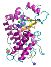
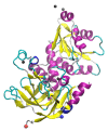
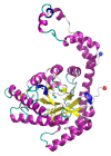
Structure of Laccase from Trametes versicolor. The crystal structure was solved by Choinowski and coworkers; downloaded from rcsb.org (1GYC) ([126,145]). The structure was visualized using Visual Molecular Dynamics (VMD) software. (A) 3D structure of laccase. The N- and C-termini are shown as red and blue spheres, respectively, while the copper ions are shown in orange (partially occluded in the structure). (B) structural geometry of the mono-copper site with chelating residues highlighted.
Figure 2
Figure 2
Structure of myoglobin from cardiac muscle of Equus caballus. The crystal structure was solved by Brayer and coworkers; downloaded from rcsb.org (1WLA) [145,173]. The structure was visualized using VMD. (A) 3D structure of myoglobin. The N- and C-termini are shown as red and blue spheres respectively while the heme is shown in orange (partially occluded in the structure). (B) Structural geometry of the heme with the iron shown in black and chelating residues highlighted.
Figure 3
Figure 3
Structure of azurin from Pseudomonas areuginosa. The crystal structure was solved by Adman and Jensen; downloaded from rcsb.org (1AZU ([182]) [145] The structure was visualized using VMD. (A) 3D structure of azurin. The N- and C-termini are shown as red and blue spheres respectively while the copper is shown in orange (partially occluded in the structure). (B) Structural geometry of the copper shown in orange and chelating residues highlighted.
Figure 4
Figure 4
3D Structure of horseradish peroxidase from Armoracia rusticana. The crystal structure was solved by Hajdu and coworkers; downloaded from rcsb.org (1W4Y) [145,186]. The structure was visualized using VMD. The N- and C-termini are shown as red and blue spheres respectively while the calcium ions are shown in green, the heme in orange and the heme-iron in black.
Figure 5
Figure 5
3D Structure of alcohol dehydrogenase from Saccharomyces cerevisiae. The crystal structure was solved by Ramaswamy and coworkers; downloaded from rcsb.org (5ENV) ([145,193]. The structure was visualized using VMD. The N- and C-termini are shown as red and blue spheres respectively while the zinc ions are shown in black. The structure represents one monomer of a homotetramer.
Figure 6
Figure 6
3D Structure of glucose isomerase from Streptomyces rubiginosus. The crystal structure was solved by Dauter and coworkers; downloaded from rcsb.org (1OAD) [145,199] The structure was visualized using VMD. The N- and C-termini are shown as red and blue spheres respectively while the manganase ions are shown in tan and the magnesium ions shown in cyan. The structure represents one monomer of a homodimer.

Similar articles

Cited by

References

    1. Branden C.I., Tooze J. Introduction to Protein Structure. Garland Science; New York, NY, USA: 2012.
    1. Lesk A.M. Introduction to Protein Architecture: The Structural Biology of Proteins. Oxford University Press; New York, NY, USA: 2001.
    1. Lesk A. Introduction to Protein Science: Architecture, Function, and Genomics. Oxford University Press; New York, NY, USA: 2010.
    1. Leader B., Baca Q.J., Golan D.E. Protein therapeutics: A summary and pharmacological classification. Nat. Rev. Drug Discov. 2008;7:21–39. doi: 10.1038/nrd2399. - DOI - PubMed
    1. Dimitrov D. Therapeutic Proteins. In: Voynov V., Caravella J.A., editors. Methods in Molecular Biology. Humana Press; Totowa, NJ, USA: 2012. pp. 1–26. - PMC - PubMed

MeSH terms

LinkOut - more resources