Review
. 2021 Jan 19;10(1):199.
doi: 10.3390/foods10010199.

Omega-3 Polyunsaturated Fatty Acids and the Intestinal Epithelium-A Review

Affiliations
Review

Omega-3 Polyunsaturated Fatty Acids and the Intestinal Epithelium-A Review

Luke A Durkin et al. Foods. .

Abstract

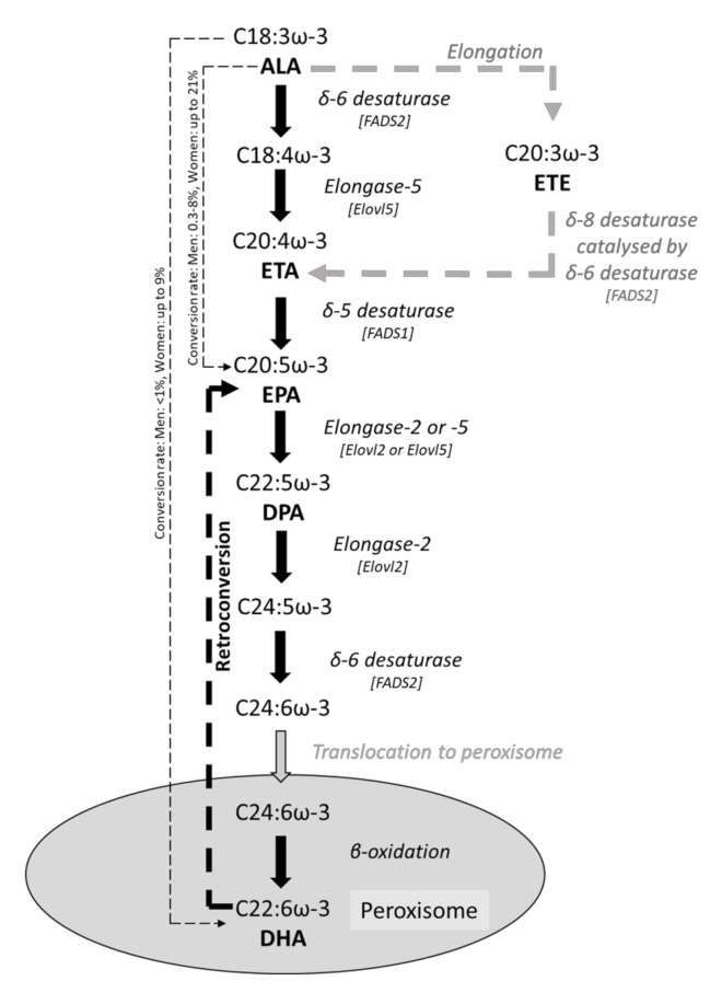
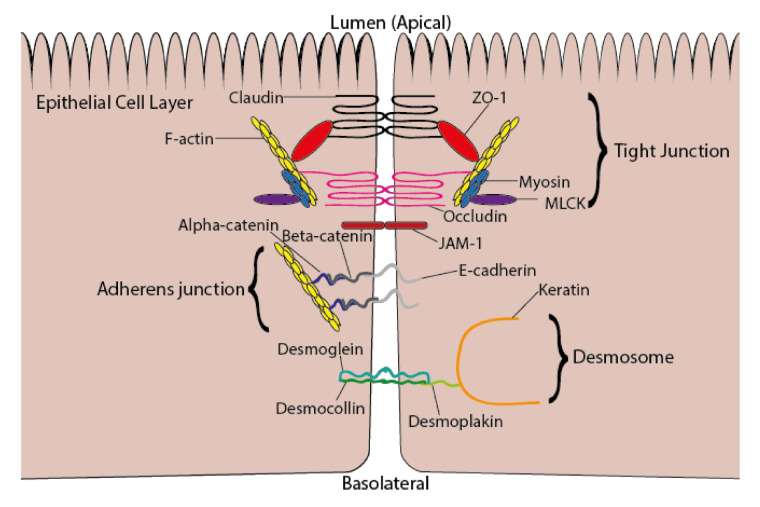
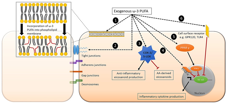
Epithelial cells (enterocytes) form part of the intestinal barrier, the largest human interface between the internal and external environments, and responsible for maintaining regulated intestinal absorption and immunological control. Under inflammatory conditions, the intestinal barrier and its component enterocytes become inflamed, leading to changes in barrier histology, permeability, and chemical mediator production. Omega-3 (ω-3) polyunsaturated fatty acids (PUFAs) can influence the inflammatory state of a range of cell types, including endothelial cells, monocytes, and macrophages. This review aims to assess the current literature detailing the effects of ω-3 PUFAs on epithelial cells. Marine-derived ω-3 PUFAs, eicosapentaenoic acid and docosahexaenoic acid, as well as plant-derived alpha-linolenic acid, are incorporated into intestinal epithelial cell membranes, prevent changes to epithelial permeability, inhibit the production of pro-inflammatory cytokines and eicosanoids and induce the production of anti-inflammatory eicosanoids and docosanoids. Altered inflammatory markers have been attributed to changes in activity and/or expression of proteins involved in inflammatory signalling including nuclear factor kappa-light-chain-enhancer of activated B cells (NF-κB), peroxisome proliferator activated receptor (PPAR) α and γ, G-protein coupled receptor (GPR) 120 and cyclooxygenase (COX)-2. Effective doses for each ω-3 PUFA are difficult to determine due to inconsistencies in dose and time of exposure between different in vitro models and between in vivo and in vitro models. Further research is needed to determine the anti-inflammatory potential of less-studied ω-3 PUFAs, including docosapentaenoic acid and stearidonic acid.

Keywords: chemokine; cytokine; eicosanoid; enterocyte; epithelium; fish oil; inflammation; lipid mediator; permeability; ω-3 PUFA.


Conflict of interest statement

L.A.D. and C.E.C. declare no conflict of interest. P.C.C. acts as a consultant to BASF AS, Smartfish, DSM, Cargill, Danone/Nutricia and Fresenius-Kabi.

Figures

Figure 1
Figure 1
Metabolic conversion pathway from the essential ω-3 PUFA, ALA, to longer-chain ω-3 PUFAs, EPA, DPA and DHA. Conversion data are from [18].
Figure 2
Figure 2
Intercellular junctions between epithelial cells consisting of tight junctions, adherens junctions, and desmosomes. Abbreviations used: ZO-1, zonula occludens-1; MLCK, myosin light chain kinase; JAM-1, junction adhesion molecule-1.
Figure 3
Figure 3
IL-8 and monokine induced by gamma interferon (MIG) production by Caco-2 cells stimulated with a cocktail of cytokines (TNF-α (5 ng/mL), IFN-γ (50 ng/mL), and IL-1β) in a Transwell system; cytokines were added to the basolateral compartment, and apical and basolateral supernatants assessed for IL-8 and MIG by Luminex. Controls are unstimulated. (a): Apical IL-8 production. (b): Basolateral IL-8 production. (c): Apical MIG production. (d): Basolateral MIG production. Data are the mean ± SEM from three experiments and are not previously published. Significance was determined by one-way ANOVA with Dunnett’s multiple comparison tests; ** p < 0.01, *** p < 0.001, and **** p < 0.0001.
Figure 4
Figure 4
Proposed mechanisms involved in ω-3 PUFA regulation of inflammation in intestinal epithelial cells. (1). Incorporation of ω-3 PUFA into phospholipid membrane/lipid rafts. (2). Modulation of tight junction protein expression and redistribution. (3). Production of anti-inflammatory eicosanoids and inhibition of AA-derived eicosanoids catalysed by COX-2 or 5-LOX. (4). Activation of nuclear receptors, e.g., PPAR-α. (5). Translocation of transcription factors into nucleus, e.g., PPAR-γ. (6). Interaction with transmembrane/cell surface receptors, e.g., G-protein coupled receptor 120 (GPR120) and TLR4. Mechanisms 4, 5, and 6 lead to the inhibition of NF-κB and the subsequent reduced production of multiple inflammatory mediators.

References

    1. McCole D.F., Barrett K.E. Varied role of the gut epithelium in mucosal homeostasis. Curr. Opin. Gastroenterol. 2007;23:647–654. doi: 10.1097/MOG.0b013e3282f0153b. - DOI - PubMed
    1. Michielan A., D’Inca R. Intestinal permeability in inflammatory bowel disease: Pathogenesis, clinical evaluation, and therapy of leaky gut. Mediat. Inflamm. 2015;2015:628157. doi: 10.1155/2015/628157. - DOI - PMC - PubMed
    1. Morrison D.J., Preston T. Formation of short chain fatty acids by the gut microbiota and their impact on human metabolism. Gut Microbes. 2016;7:189–200. doi: 10.1080/19490976.2015.1134082. - DOI - PMC - PubMed
    1. Shi N., Li N., Duan X., Niu H. Interaction between the gut microbiome and mucosal immune system. Milit. Med. Res. 2017;4:14. doi: 10.1186/s40779-017-0122-9. - DOI - PMC - PubMed
    1. Guillot X., Semerano L., Saidenberg-Kermanac’h N., Falgarone G., Boissier M.-C. Vitamin D and inflammation. Joint Bone Spine. 2010;77:552–557. doi: 10.1016/j.jbspin.2010.09.018. - DOI - PubMed