. 2021 Jan 19;13(2):313.
doi: 10.3390/polym13020313.

Deciphering Structural Determinants in Chondroitin Sulfate Binding to FGF-2: Paving the Way to Enhanced Predictability of their Biological Functions

Affiliations

Deciphering Structural Determinants in Chondroitin Sulfate Binding to FGF-2: Paving the Way to Enhanced Predictability of their Biological Functions

Giulia Vessella et al. Polymers (Basel). .

Abstract

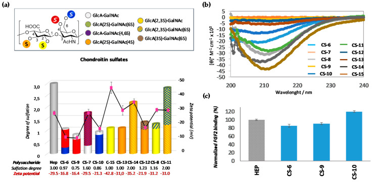
Controlling chondroitin sulfates (CSs) biological functions to exploit their interesting potential biomedical applications requires a comprehensive understanding of how the specific sulfate distribution along the polysaccharide backbone can impact in their biological activities, a still challenging issue. To this aim, herein, we have applied an "holistic approach" recently developed by us to look globally how a specific sulfate distribution within CS disaccharide epitopes can direct the binding of these polysaccharides to growth factors. To do this, we have analyzed several polysaccharides of marine origin and semi-synthetic polysaccharides, the latter to isolate the structure-activity relationships of their rare, and even unnatural, sulfated disaccharide epitopes. SPR studies revealed that all the tested polysaccharides bind to FGF-2 (with exception of CS-8, CS-12 and CS-13) according to a model in which the CSs first form a weak complex with the protein, which is followed by maturation to tight binding with k D ranging affinities from ~ 1.31 μM to 130 μM for the first step and from ~ 3.88 μM to 1.8 nM for the second one. These binding capacities are, interestingly, related with the surface charge of the 3D-structure that is modulated by the particular sulfate distribution within the disaccharide repeating-units.

Keywords: chondroitin sulfate; fibroblast growth factor 2; glycosaminoglycan; protein interactions; structure-activity relationships.


Conflict of interest statement

The authors declare no conflict of interest.

Figures

Figure 1
Figure 1
(a) Structure of chondroitin sulfate chains (top); chemical structure of typical disaccharide units found in CS chains (bottom). An “S” enclosed by circles indicates the various positions that can be esterified by sulfates. (b) Disaccharide sulfation patterns of natural CSs.
Figure 2
Figure 2
Schematic diagram of our proposal.
Figure 3
Figure 3
Scheme of the semi-synthesis of CS-7.
Figure 4
Figure 4
Scheme of the synthesis of CS-8, CS-11 and CS-12.
Figure 5
Figure 5
Schematic representation of the polysaccharides systematically analyzed in this study. Natural CSs are highlighted with a grey background. The grey dashed lines indicate differences in terms of disaccharide motifs among natural CSs. These motifs were isolated in the polysaccharides that are highlighted with a green background. Finally, a red background indicates polysaccharides found to recapitulate rare or unnatural sulfation patterns in GlcA. The differences between these polysaccharides are shown over the red dashed arrows.
Figure 6
Figure 6
1H and DEPT-HSQC NMR spectra (400 MHz, D2O, 298 K, zoom) of CS-7 (a), CS-8 (b), CS-11 (c), and CS-12 (d). In Figure 6a, GalNAc is referred to both 4,6-disulfated and zero-sulfated GalNAc units. In Figure 6b–d, GalNAc are referred to both 6-sulfated and zero-sulfated GalNAc units. Densities enclosed in the highlighted areas were integrated for disaccharide residue ratio estimation. For full 1H and DEPT-HSQC NMR spectra, see Supporting Information (Figures S1–S4).
Figure 7
Figure 7
(a) Degree of sulfation of polysaccharides (their numerical values are indicated at the bottom in grey). The pink points indicate the zeta-potential values (their numerical values are indicated at the bottom, in red). The sulfated-disaccharide composition of each polysaccharide is shown in colours (for interpretation, see figure legend). (b) CD spectra of polysaccharides. (c) Bar graphs of normalized FGF-2 binding. The concentrations of polysaccharides were 0.5 mg/mL. All bar graphs are based on triplicate experiments. Heparine (Hep) was employed as reference.
Figure 8
Figure 8
(a) Degree of sulfation of polysaccharides (their numerical values are indicated at the bottom, in grey). The pink points indicate the zeta-potential values (their numerical values are indicated at the bottom, in red). Sulfated-disaccharide composition of each polysaccharide is shown in colors (for interpretation, see figure legend). (b) CD spectra of polysaccharides. (c) SPR binding of immobilized FGF-2 to GalNAc sulfated derivatives (CS-6, CS-9 and CS-10) (0.5 mg/mL) normalized against heparin. All bar graphs are based on triplicate experiments.
Figure 9
Figure 9
(a) Bar graph of normalized CS-7 binding preference to surface FGF-2 by competing with different concentrations of CS-15. The dashed red line indicates the lever of responsiveness of CS-7 alone. (b) SPR sensograms for binding-affinity measurements. The concentration of CS-7 was, in all cases, 10 µM, and concentrations of CS-15 were (from top to bottom): 50 µM, 20 µM, 10 µM and 0 µM.
Figure 10
Figure 10
Dual function of chondroitin sulfates in their binding to FGF-2 as mediators of growth factor signaling to cells (right) or as providers of storage sites, protecting and stabilization the protein (left).

References

    1. Bedini E., Laezza A., Iadonisi A. Chemical Derivatization of Sulfated Glycosaminoglycans. Eur. J. Org. Chem. 2016;18:3018–3043. doi: 10.1002/ejoc.201600108. - DOI
    1. López-Álvarez M., López-Senra E., Valcárcel J., Vázquez J.A., Serra J., González P. Quantitative Evaluation of Sulfation Position Prevalence in Chondroitin Sulphate by Raman Spectroscopy. J. Raman Spectrosc. 2019;50:656–664. doi: 10.1002/jrs.5563. - DOI
    1. Bedini E., Corsaro M.M., Fernández-Mayoralas A., Iadonisi A. Chondroitin, Dermatan, Heparan, and Keratan Sulfate: Structure and Functions. In: Cohen E., Merzendorfer H., editors. Extracellular Sugar-Based Biopolymers Matrices. Biologically-Inspired Systems. 1st ed. Volume 12. Springer; Cham, Switzerland: 2019. pp. 187–233.
    1. Kjellén L., Lindahl U. Specificity of Glycosaminoglycan-protein Interactions. Curr. Opin. Struct. Biol. 2018;50:101–108. doi: 10.1016/j.sbi.2017.12.011. - DOI - PubMed
    1. Gama C.I., Tully S.E., Sotogaku N., Clark P.M., Rawat M., Vaidehi N., Goddard W.A., Nishi A., Hsieh-Wilson L.C. Sulfation Patterns of Glycosaminoglycans Encode Molecular Recognition and Activity. Nat. Chem. Biol. 2006;2:467–473. doi: 10.1038/nchembio810. - DOI - PubMed

LinkOut - more resources