. 2021 Jan 21;21(1):54.
doi: 10.1186/s12870-021-02833-w.

An improved and efficient method of Agrobacterium syringe infiltration for transient transformation and its application in the elucidation of gene function in poplar

Affiliations

An improved and efficient method of Agrobacterium syringe infiltration for transient transformation and its application in the elucidation of gene function in poplar

Lin Zheng et al. BMC Plant Biol. .

Abstract

Background: Forest trees have important economic and ecological value. As a model tree, poplar has played a significant role in elucidating the molecular mechanisms underlying tree biology. However, a lack of mutant libraries and time-consuming stable genetic transformation processes severely limit progress into the functional characterization of poplar genes. A convenient and fast transient transformation method is therefore needed to enhance progress on functional genomics in poplar.

Methods: A total of 11 poplar clones were screened for amenability to syringe infiltration. Syringe infiltration was performed on the lower side of the leaves of young soil-grown plants. Transient expression was evaluated by visualizing the reporters β-glucuronidase (GUS) and green fluorescent protein (GFP). The experimental parameters of the syringe agroinfiltration were optimized based on the expression levels of the reporter luciferase (LUC). Stably transformed plants were regenerated from transiently transformed leaf explants through callus-induced organogenesis. The functions of Populus genes in secondary cell wall-thickening were characterized by visualizing lignin deposition therein after staining with basic fuchsin.

Results: We greatly improved the transient transformation efficiency of syringe Agrobacterium infiltration in poplar through screening for a suitable poplar clone from a variety of clones and optimizing the syringe infiltration procedure. The selected poplar clone, Populus davidiana × P. bolleana, is amenable to Agrobacterium syringe infiltration, as indicated by the easy diffusion of the bacterial suspension inside the leaf tissues. Using this technique, we localized a variety of poplar proteins in specific intracellular organelles and illustrated the protein-protein and protein-DNA interactions. The transiently transformed leaves could be used to generate stably transformed plants with high efficiency through callus induction and differentiation processes. Furthermore, transdifferentiation of the protoxylem-like vessel element and ectopic secondary wall thickening were induced in the agroinfiltrated leaves via the transient overexpression of genes associated with secondary wall formation.

Conclusions: The application of P. davidiana × P. bolleana in Agrobacterium syringe infiltration provides a foundation for the rapid and high-throughput functional characterization of Populus genes in intact poplar plants, including those involved in wood formation, and provides an effective alternative to Populus stable genetic transformation.

Keywords: Poplar; Secondary wall formation; Syringe Agrobacterium infiltration; Transgenic poplar; Transient expression.


Conflict of interest statement

The authors declare that they have no competing interests.

Figures
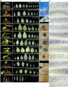
Fig. 1
Fig. 1
The spreadability of the agrobacterial suspension and expression of reporters in the tested poplar clones. The transformations were carried out using A. tumefaciens EHA105, which was suspended in the infiltrated medium [10 mM MgCl2, 5 mM MES-KOH (pH 5.6) and 0.2 mM Acetosyringone (AS)]. a The spreadability of the agrobacterial suspension in the leaves of the tested poplar clones. Agroinfiltration was performed on leaves from leaf Plastochron index (LPI) 1 down to the last one (shown sequentially from left to right). The bacterial suspension spread well in clones P. davidiana × bolleana, P. alba var. pyramidalis, and P. trichocarpa, with the best performance observed in the leaves LPI 4 of P. davidiana × bolleana, LPI 4 of P. alba var. pyramidalis, and LPI 3 of P. trichocarpa, as indicated by the red stars. In contrast, the bacterial suspension was shown to be limited to a very small region in all the manipulated leaves in the other clones. The suspension was clearly delineated by leaf veins in leaves LPI 3 of clones P. tomentosa ‘B331’ and P. popularis ‘35–44’. Bars = 2 cm. b The GUS staining in the leaves of the tested clones. All infiltrated leaves were stained, and the representative images are shown. Bars = 2 mm. c The interior structure of the full-expanded leaves of the tested clones. Five-micrometer-thick sections were stained with TBO and observed using a Leica DM 5500 B light microscope. The clones P. davidiana × bolleana, P. alba var. pyramidalis, and P. trichocarpa showed larger intercellular air spaces inside the leaves compared to the other clones, in which the air spaces were smaller and more compartmented. The mesophyll cells were arranged randomly and loosely within the leaves in the clone P. davidiana × bolleana
Fig. 2
Fig. 2
Factors affecting the transient expression efficiency in poplar leaves. Optimization of the experimental parameters was carried out on P. davidiana × bolleana. a Effect of the plant development stage on transient expression efficiency. Syringe agroinfiltration was conducted on leaves LPI 4 using A. tumefaciens GV3101 suspended in modified infiltration medium [10 mM MgCl2, 5 mM MES-KOH (pH 5.6) and 1.6 mM AS] and evaluated at 5 dpi. b Effect of leaf age on transient expression efficiency. The syringe agroinfiltration was conducted on plants PI 11–12 using GV3101 suspended in modified infiltration medium [10 mM MgCl2, 5 mM MES-KOH (pH 5.6) and 1.6 mM AS] and evaluated at 5 dpi. c Effect of Agrobacterium strains on transient expression efficiency. The syringe agroinfiltration was conducted on the leaves LPI 4 of plants PI 11–12 by using the indicated A. tumefaciens strains suspended in modified infiltration medium [10 mM MgCl2, 5 mM MES-KOH (pH 5.6) and 1.6 mM AS] and evaluated at 5 dpi. d Effect of acetosyringone concentration on transient expression efficiency. The syringe agroinfiltration was carried out on leaves LPI 4 of plants PI 11–12 by using GV3101 suspended in the infiltration media [10 mM MgCl2, 5 mM MES-KOH (pH 5.6)] supplemented with different concentrations of AS and evaluated at 5 dpi. e Effect of the duration of transient expression on the transient expression efficiency. The syringe agroinfiltration was carried on leaves LPI 4 of plants PI 11–12 by using GV3101 suspended in modified infiltration medium [10 mM MgCl2, 5 mM MES-KOH (pH 5.6) and 1.6 mM AS]. LUC activity was evaluated at 1, 3, 5, 7, 9, and 11 dpi. The different letters above the bar indicate statistically significant differences, while the same letter indicates no significant difference according to Duncan’s (D) test (P < 0.05). The red line shows the average LUC activity (n = 8)
Fig. 3
Fig. 3
The ideal P. davidiana ×bolleana plant for agroinfiltration and transient expression of reporter gene. a The ideal plant with an age of PI 12 and its optimum leaves of LPI 4 and LPI 5 used for agroinfiltration. The syringe agroinfiltration was conducted using A. tumefaciens GV3101 suspended in modified infiltration medium [10 mM MgCl2, 5 mM MES-KOH (pH 5.6) and 1.6 mM AS]. b Transient expression of green fluorescent protein (GFP) reporter in epidermal cells and mesophyll cells. The expression of GFP was evaluated at 5 dpi. To observe the fluorescent signals in the mesophyll cells, the lower epidermis was removed with a tweezer. The upper two rows show GFP expression in the epidermal cells with different magnitudes of enlargement. The third row shows GFP expression in the mesophyll cells
Fig. 4
Fig. 4
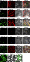
Subcellular localization of various poplar proteins. The various GFP-fused poplar proteins were driven by the Super promoter. The A. tumefaciens GV3101 suspension was infiltrated into the leaves of P. davidiana × bolleana plants under the optimal experimental parameters described above in the Results section. At 5 dpi, the infiltrated leaves were detached, and GFP fluorescent signals were observed under a Nikon inverted fluorescence microscope TE2000-E with excitation at 488 nm and emission at 510 nm. a PdbCBL1-GFP localized in the plasma membrane, consistent with FM4–64 staining. b PdbMTP1-GFP localized in the tonoplast, distinguished from the FM4–64-stained plasma membrane indicated by white arrows. c Colocalization of PdbC4H-GFP in the endoplasmic reticulum (ER) with ER marker HDEL-mCherry. d Colocalization of PdbGT47C-GFP in the Golgi with Golgi marker NAG-mCherry. e Localization of PtoMYB221-GFP within the nucleus, consistent with DAPI staining. f Localization of PdbPrxQ-GFP within plastids, consistent with chlorophyll autofluorescence. g Localization of GFP driven under the Super promoter in the cytoplasm and the nucleus
Fig. 5
Fig. 5
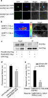
Protein–protein and protein–DNA interactions in the cells of the poplar leaves. Protein–protein interactions were illustrated with various methods using transient co-transformation in the poplar leaves by syringe infiltration under the optimal experimental parameters described above in the Results section. a. BiFC assay of AtWRKY40-YFPN and AtWRKY40-YFPC. The combination of AtWRKY40-YFPN and YFPC was used as a negative control. At 5 dpi, the infiltrated leaves were detached, and the YFP fluorescent signals were observed under a Nikon inverted fluorescence microscope TE2000-E. YFP signals and DAPI fluorescence overlapped in the nucleus. b. Split luciferase assay of AtNRT3.1-Nluc and AtNRT2.1-Cluc, showing stronger LUC activity compared to the negative control combination of AtNRT3.1-Nluc and Cluc. At 5 dpi, the transformed leaves were infiltrated with 2 mM luciferin by using a syringe without a needle, left in dark for 6 min to quench the fluorescence, and then detached for the luminescence intensity assay. The color scale shows the luminescence intensity, with blue indicating the lowest and red the highest. c. Images of the co-immunoprecipitation assay show the interaction of PtoUBC34s with PtoMYB221. PtoUBC34s-Flag was co-expressed with PtoMYB221-Myc, and protein extracts were incubated with anti-Flag coupled agarose. Immunoprecipitates (IP) and input proteins were analyzed by immunoblotting using anti-Flag and anti-Myc antibodies. The uncropped images can be found in Fig. S7. d. In vivo Förster resonance energy transfer (FRET-FLIM) measurement of co-expressing YFP-PtoUBC34s and PtoMYB221-RFP, with YFP-PtoUBC34s as the donor. The YFP-PtoUBC34s donor alone used as a negative control. The data are presented as means ±SE (n = 3). *P < 0.05, Student’s t-test. e. Dual LUC assay detected the repression ability of the EAR-like motif repression domain of SUPERMAN (SUPRD) [48, 54]. Relative LUC activities were measured after co-transformation with the reporter and effectors, where pGreen-SK was used as a control vector. The data are presented as means ±SE (n = 6). ** P < 0.01, Student’s t-test
Fig. 6
Fig. 6
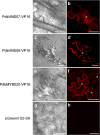
Induction of protoxylem tracheary element differentiation and secondary wall deposition in the epidermal cells. Transient overexpression of PdbVNS07 (VND-, NST/SND- and SMB-related protein), PdbVNS09 or PdbMYB020 fused with the activation domain of the herpes virus VP16 protein in poplar leaves via syringe agroinfiltration resulted in ectopic secondary wall deposition in the epidermal cells. Transiently transformed leaves were detached, stained with basic fuchsin at 10 dpi, and observed with a confocal microscope for secondary walls. a and b. Epidermal cells overexpressing PdbVNS07-VP16, showing transdifferentiation of protoxylem-like vessel elements with annular and spiral thickenings in the cell wall. c and d. Epidermal cells overexpressing PdbVNS09-VP16, showing the ectopic secondary wall deposition. e and f. Epidermal cells overexpressing PdbMYB020-VP16, showing band-like secondary wall thickening. g and h. Epidermal cells overexpressing the control vector pGreenII 62-SK, showing no secondary wall thickening. a, c, e, and g are images of differential interference contrast; b, d, f, and h are images of the basic fuchsin stain. White stars indicated ectopic secondary wall deposition

References

    1. Bradshaw H, Ceulemans R, Davis J, Stettler R. Emerging model systems in plant biology: poplar (Populus) as a model forest tree. J Plant Growth Regul. 2000;19(3):306–313. doi: 10.1007/s003440000030. - DOI
    1. Wullschleger SD, Jansson S, Taylor G. Genomics and forest biology: Populus emerges as the perennial favorite. Plant Cell. 2002;14(11):2651–2655. doi: 10.1105/tpc.141120. - DOI - PMC - PubMed
    1. Ma J, Wan D, Duan B, Bai X, Bai Q, Chen N, et al. Genome sequence and genetic transformation of a widely distributed and cultivated poplar. Plant Biotechnol J. 2019;17(2):451–460. doi: 10.1111/pbi.12989. - DOI - PMC - PubMed
    1. Tuskan GA, Difazio S, Jansson S, Bohlmann J, Grigoriev I, Hellsten U, et al. The genome of black cottonwood, Populus trichocarpa (Torr. & gray) Science. 2006;313(5793):1596–1604. doi: 10.1126/science.1128691. - DOI - PubMed
    1. Yang Y, Li R, Qi M. In vivo analysis of plant promoters and transcription factors by agroinfiltration of tobacco leaves. Plant J. 2000;22(6):543–551. doi: 10.1046/j.1365-313x.2000.00760.x. - DOI - PubMed

LinkOut - more resources