Review
. 2021 Jan 21;22(1):96.
doi: 10.1186/s12891-021-03967-6.

An unusual example of hereditary multiple exostoses: a case report and review of the literature

Affiliations
Review

An unusual example of hereditary multiple exostoses: a case report and review of the literature

Rebecca Chilvers et al. BMC Musculoskelet Disord. .

Abstract

Background: Hereditary multiple exostoses (HME) is a rare skeletal disorder characterised by a widespread. distribution of osteochondromas originating from the metaphyses of long bones.

Case presentation: This case study examines a 55-year-old male cadaver bequeathed to the University of Liverpool who suffered from HME, thus providing an exceptionally rare opportunity to examine the anatomical changes associated with this condition.

Conclusions: Findings from imaging and dissection indicated that this was a severe case of HME in terms of the quantity and distribution of the osteochondromas and the number of synostoses present. In addition, the existence of enchondromas and the appearance of gaps within the trabeculae of affected bones make this a remarkable case. This study provides a comprehensive overview of the morbidity of the disease as well as adding to the growing evidence that diseases concerning benign cartilaginous tumours may be part of a spectrum rather than distinct entities.

Keywords: Diaphyseal aclasis; Enchondroma; Hereditary multiple Exostoses; Metachondromatosis; Osteochondroma; Synostosis.


Conflict of interest statement

The authors declare that they have no competing interests.

Figures

Fig. 1
Fig. 1
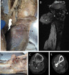
Right proximal forearm. a Superficial dissection. A tumour has caused lateral displacement of the brachioradialis muscle (a). The radial nerve (c) has been diverted over the superior surface of radial head (b). b Deep dissection. Exostosis in the region of the radial tuberosity (a), radial head dislocation (b). The capitulum (c) formed an articulation with the radius at the radial tuberosity (a), causing the biceps brachii tendon (d) to be incorporated into the joint. c DESS MRI of proximal radius. An osteochondroma present at the radial tuberosity (a) has a continuous cortex and medulla with the underlying bone. A cartilage cap is present on the surface (b). Note the low signal within the head of the radius (c). d Sagittal section of radial head. Radius displays a large void (a) within the epiphysis
Fig. 2
Fig. 2
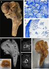
Right proximal femur. a Posterior view. A large exostosis (a) surrounding the metaphysis with an outgrowth (b) from the lateral portion of the femoral shaft. Greater trochanter (c) and femoral head also displayed (d). b A section through intramedullary mass. (a) Cartilage matrix (b) inter nodular cartilage. c A section through the surface of a sessile osteochondroma. (a) Perichondrium on surface of cartilage cap. (b) chondrocytes. (c) Hypertrophic mature chondrocytes. (d) Subchondral bone. d T2 MRI coronal section. Osteochondromas can be seen surrounding the metaphysis and extending from the diaphysis (a and b respectively). Cartilage caps can also be seen on the osteochondromas (c). A large fluid filled mass is present in the metaphysis (d). Large voids are present in both the epiphysis and diaphysis (e and f). e Transverse CT scan through greater trochanter (a). Showing osteochondroma (b). f Transverse CT scan through proximal diaphysis. The pedunculated osteochondroma (a) can be seen on the lateral surface whilst the sessile growth can be seen on the posterior diaphysis (b). g Coronal section. (a) Cross section of osteochondroma. (b) trabecular bone obliterating normal cortical bone as the medulla of the overlying exostosis. (c) Cortex of exostosis. (d) Soft mass located in the metaphysis. (e) An example of the gaps located within the trabecular bone. h Cartilaginous mass removed (a) surrounded by trabeculae (b)
Fig. 3
Fig. 3
Right proximal leg. a Superficial dissection. Gastrocnemius (a) has been reflected to show the osteochondroma (b) of the head of the fibula causing deformity of the soleus muscle (c). b Lateral view. (a) Fibula. (b) Cartilage capped osteochondromas of head of fibula. c Transverse CT scan through fibular head. Osteochondroma visible with cortical bone (a) and trabecular (b) continuous with the underlying normal bone. A synostosis between the tibia and fibula can be seen (c). d DESS MRI sagittal section. The osteochondroma of the fibular head can be seen containing trabecular bone (a). The synostosis between the fibular and tibia heads can be observed (b). Like the femur and radius the tibia contains large gaps in the trabecular bone structure (c). Cortical destruction has occurred (d). e Transverse CT scan through distal tibia and fibula. An osteochondroma on either the tibia (a) or fibular (b) has formed a synostosis (c) at the distal end of the two bones
Fig. 4
Fig. 4
Right foot. a Radiograph. A pedunculated growth is present on the medial surface of the 1st distal phalanx (a). A sessile osteochondroma is situated on the inferior surface of the 1st metatarsal bone (b). A sysnostosis has formed between the heads of the 2nd and 3rd metatarsals (c). An osteochondroma of the 2nd metatarsal has altered the orientation of the metatarsophalangeal joint (d). b Superficial dissection. The cartilage capped osteochondroma can be seen at the head on the 2nd metatarsal (a). A tendon of extensor digitorum longus is observed inserting into the osteochondroma (b)

References

    1. Upadhyaya GK, Jain VK, Arya RK, Sinha S, Naik AK. Osteochondroma of upper dorsal spine causing spastic Paraparesis in hereditary multiple Exostosis: a case report. J Clin Diagn Res. 2015;9(12):RD04–RD06. - PMC - PubMed
    1. Beltrami G, Ristori G, Scoccianti G, Tamburini A, Capanna R. Hereditary multiple Exostoses: a review of clinical appearance and metabolic pattern. Clin Cases Miner Bone Metab. 2016;13(2):110–118. - PMC - PubMed
    1. DuBose CO. Multiple Hereditary Exostoses. Radiol Technol. 2016;87(3):305–321. - PubMed
    1. Sajid S, Yousaf A, Nabi U, Shahbaz A, Amin U. Sarcomatous transformation of recurrent scapular Osteochondroma in a patient with the hereditary multiple Osteochondromas: a case report and literature review. Cureus. 2019;11(12):e6308. - PMC - PubMed
    1. Garcia-Gonzalez O, Mireles-Cano JN, Sanchez-Zavala N, Chagolla-Santillan MA, Orozco-Ramirez SM, Silva-Cerecedo P, Murguia-Perez M, Rueda-Franco F. Multiple hereditary osteochondromatosis with spinal cord compression: case report. Childs Nerv Syst. 2018;34(3):565–569. doi: 10.1007/s00381-017-3645-1. - DOI - PubMed