. 2021 Jan 21;9(1):18.
doi: 10.1186/s40168-020-00965-z.

Decoding the RNA viromes in rodent lungs provides new insight into the origin and evolutionary patterns of rodent-borne pathogens in Mainland Southeast Asia

Affiliations

Decoding the RNA viromes in rodent lungs provides new insight into the origin and evolutionary patterns of rodent-borne pathogens in Mainland Southeast Asia

Zhiqiang Wu et al. Microbiome. .

Abstract

Background: As the largest group of mammalian species, which are also widely distributed all over the world, rodents are the natural reservoirs for many diverse zoonotic viruses. A comprehensive understanding of the core virome of diverse rodents should therefore assist in efforts to reduce the risk of future emergence or re-emergence of rodent-borne zoonotic pathogens.

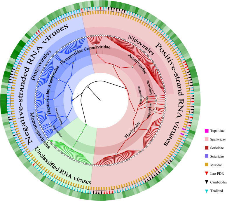
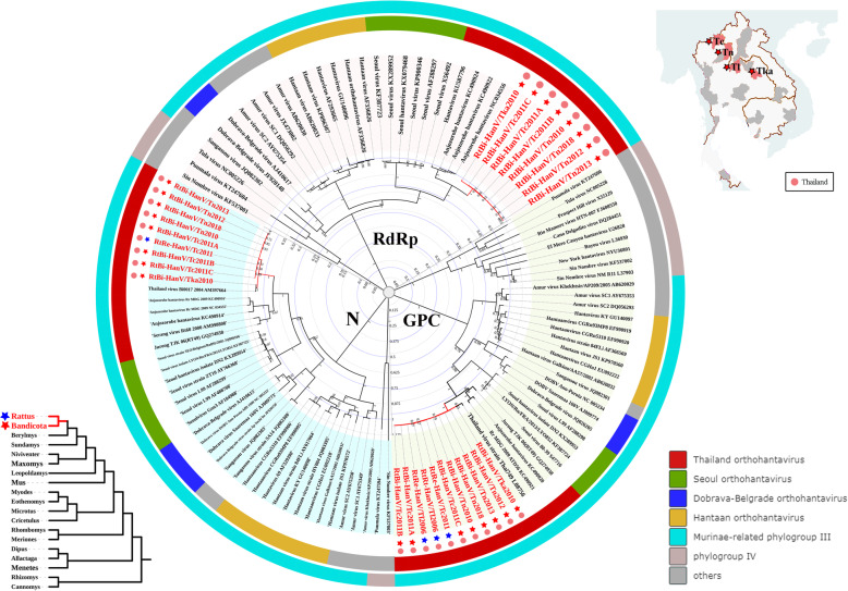
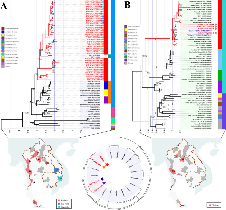
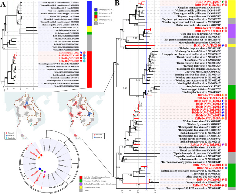
Results: This study aimed to describe the viral range that could be detected in the lungs of rodents from Mainland Southeast Asia. Lung samples were collected from 3284 rodents and insectivores of the orders Rodentia, Scandentia, and Eulipotyphla in eighteen provinces of Thailand, Lao PDR, and Cambodia throughout 2006-2018. Meta-transcriptomic analysis was used to outline the unique spectral characteristics of the mammalian viruses within these lungs and the ecological and genetic imprints of the novel viruses. Many mammalian- or arthropod-related viruses from distinct evolutionary lineages were reported for the first time in these species, and viruses related to known pathogens were characterized for their genomic and evolutionary characteristics, host species, and locations.

Conclusions: These results expand our understanding of the core viromes of rodents and insectivores from Mainland Southeast Asia and suggest that a high diversity of viruses remains to be found in rodent species of this area. These findings, combined with our previous virome data from China, increase our knowledge of the viral community in wildlife and arthropod vectors in emerging disease hotspots of East and Southeast Asia. Video abstract.

Keywords: Core virome; Emerging infectious diseases; Mainland Southeast Asia; Rodent lungs; Viral evolution.


Conflict of interest statement

The authors declare that they have no competing interests

Figures

Fig. 1
Fig. 1
a Sampling sites from the various provinces of Southeast Asia. Map of Southeast Asia showing the eighteen provinces in the three countries where animals were sampled. Sampling sites are indicated in different color, with red color representing Thailand, blue representing Lao PDR, and cyan representing Cambodia. Sampling sites within the eighteen provinces are shown on the right, with dots indicating sampling sites with each province. The number of samples from the species and the provinces and dates of collection are detailed in Table S1. b Heatmap based on the normalized numbers of sequence reads for 24 families of mammalian viruses and a group of unclassified RNA viruses in each pooled sample. Mammalian host species are listed on the right as a text column. Location information is indicated on the left as color code, with colors defined on the right. Names of the mammalian viral families are indicated at the bottom. Relative abundance of the viruses in each species in each location are indicated as a heat map ranging from low (yellow) to high (red) based on the normalized average viral genome size and total sequencing reads in each pool
Fig. 2
Fig. 2
Overview of the classification, from family to order, of RNA viruses identified in this study. Different families and unclassified RNA viruses are labeled in different colors in the pie chart. The three outer rings display host mammalian family (inner ring), geographical location (middle ring), and relative abundance of virus, with light to dark green representing increasing abundance (outer ring)
Fig. 3
Fig. 3
Phylogenetic trees based on the complete M segment-encoded glycoprotein precursor (GPC), L segment-encoded RdRp (RdRp), and S segment-encoded nucleocapsid protein (N) amino acid sequences of HanVs. Phylogenetic trees were constructed by the maximum likelihood method using the best-fit models (LG + G for GPC protein, LG + G + I for RdRp protein, and GTR + G + I for N protein). All HanVs found in this study are labeled in red. Host genus and location of each virus are labeled by the 5-point stars and dots of different colors. The outer color rings represent additional taxonomic information about these viruses. DOBV, Dobrava-Belgrade virus
Fig. 4
Fig. 4
Phylogenetic tree based on the partial L (RdRP) nucleotide sequences of PhleVs (2392 bp). Phylogenetic tree was constructed by the maximum likelihood method using the best-fit model (T92 + G + I). All PhleVs found in this study are labeled in red. Host genus and location of each virus are labeled by the 5-point stars and dots of different colors. The outer color rings represent additional taxonomic information about these viruses. SFTS virus, severe fever with thrombocytopenia syndrome virus
Fig. 5
Fig. 5
Phylogenetic trees based on the complete L protein (RdRP), glycoprotein (G), and nucleocapsid (N) protein amino acid sequences of AreVs. Phylogenetic trees were constructed by the maximum likelihood method using the best-fit models (LG + G + I for L protein, GTR + G for G protein, and T92 + G for N protein). All AreVs found in this study are labeled in red. Host genus and location of each virus are labeled by the 5-point stars and dots of different colors. The outer color rings represent additional taxonomic information about these viruses. JUNV, Junin virus; PICV, Pichinde virus; DANV, Dandenong virus; LCMV, lymphocytic choriomeningitis virus; LUJV, Lujo virus; IPPYV, Ippy virus; LASV, lassa virus; MOBV, Mobala virus; MORV, Morogoro virus; MOPV, Mopeia virus
Fig. 6
Fig. 6
a Phylogenetic tree based on complete L (RdRp) nucleotide sequences of RhaV (6356 bp). b Phylogenetic tree based on partial L (RdRp) nucleotide sequences of ParaVs (559 bp). Phylogenetic trees were constructed by the maximum likelihood method using the best-fit models (GTR + G + I for RhaV and ParaV). All viruses found in this study are labeled in red. Host genus and location of each virus are labeled by the 5-point stars and dots of different colors. The outer color rings represent additional taxonomic information about these viruses. GSqRV, Giant squirrel respirovirus
Fig. 7
Fig. 7
Phylogenetic trees based on the polyprotein sequences of hepaciviruses, pegiviruses, and pestivirus (hepaciviruses and pegiviruses, 2060 bp; pestivirus, 796 aa). Phylogenetic trees were constructed by the maximum likelihood method using the best-fit models (GTR + G for hepacivirus and pegivirus, rtREV+G for pestivirus). All flaviviruses found in this study are labeled in red. Host genus and location of each virus are labeled by the 5-point stars and dots of different colors. The outer color rings represent additional taxonomic information about these viruses. APPV, atypical porcine pestivirus; LV, Linda virus; BVDV, bovine viral diarrhea virus; CFSV, classical swine fever virus; BDV, border disease virus
Fig. 8
Fig. 8
a Phylogenetic tree based on complete ORF1b nucleotide sequences of ArteVs. b Phylogenetic tree based on complete RdRp nucleotide sequences of CoVs. Phylogenetic trees were constructed by the maximum likelihood method using the best-fit models (GTR + G for ArteV, and GTR + G + I for CoV). All viruses found in this study are labeled in red. Host genus and location of each virus are labeled by the 5-point stars and dots of different colors. The outer color rings represent additional taxonomic information about these viruses. SWBV, southwest baboon virus; MYBV, Mikumi yellow baboon virus; APRAV, African pouched rat arterivirus; WPDV, Wobbly possum disease virus
Fig. 9
Fig. 9
a Phylogenetic tree based on partial ORF1 nucleotide sequences of HEVs (4392 bp). b Phylogenetic tree based on partial RdRp nucleotide sequences of unclassified RNA viruses (1014 bp). Phylogenetic trees were constructed by the maximum likelihood method using the best-fit models (GTR + G for HEV, and GTR + G for unclassified RNA viruses). All viruses found in this study are labeled in red. Host genus and location of each virus are labeled by the 5-point stars and dots of different colors. The outer color rings represent additional taxonomic information about these viruses

References

    1. Ren LL, Wang YM, Wu ZQ, Xiang ZC, Guo L, Xu T, Jiang YZ, Xiong Y, Li YJ, Li XW, et al. Identification of a novel coronavirus causing severe pneumonia in human: a descriptive study. Chin Med J (Engl) 2020;133:1015–1024. doi: 10.1097/CM9.0000000000000722. - DOI - PMC - PubMed
    1. Leroy EM, Kumulungui B, Pourrut X, Rouquet P, Hassanin A, Yaba P, Delicat A, Paweska JT, Gonzalez JP, Swanepoel R. Fruit bats as reservoirs of Ebola virus. Nature. 2005;438:575–576. doi: 10.1038/438575a. - DOI - PubMed
    1. Zhou P, Yang XL, Wang XG, Hu B, Zhang L, Zhang W, Si HR, Zhu Y, Li B, Huang CL, et al. A pneumonia outbreak associated with a new coronavirus of probable bat origin. Nature. 2020;579:270–3. - PMC - PubMed
    1. Wu F, Zhao S, Yu B, Chen YM, Wang W, Song ZG, Hu Y, Tao ZW, Tian JH, Pei YY, et al. A new coronavirus associated with human respiratory disease in China. Nature. 2020;579:265–269. doi: 10.1038/s41586-020-2008-3. - DOI - PMC - PubMed
    1. Blanga-Kanfi S, Miranda H, Penn O, Pupko T, DeBry RW, Huchon D. Rodent phylogeny revised: analysis of six nuclear genes from all major rodent clades. BMC Evol Biol. 2009;9:71. doi: 10.1186/1471-2148-9-71. - DOI - PMC - PubMed

Publication types

LinkOut - more resources