. 2021 Apr;27(4):411-419.
doi: 10.1261/rna.077602.120. Epub 2021 Jan 21.

Dynamic association of human Ebp1 with the ribosome

Affiliations

Dynamic association of human Ebp1 with the ribosome

Varun Bhaskar et al. RNA. 2021 Apr.

Abstract

Ribosomes are the macromolecular machines at the heart of protein synthesis; however, their function can be modulated by a variety of additional protein factors that directly interact with them. Here, we report the cryo-EM structure of human Ebp1 (p48 isoform) bound to the human 80S ribosome at 3.3 Å resolution. Ebp1 binds in the vicinity of the peptide exit tunnel on the 80S ribosome, and this binding is enhanced upon puromycin-mediated translational inhibition. The association of Ebp1 with the 80S ribosome centers around its interaction with ribosomal proteins eL19 and uL23 and the 28S rRNA. Further analysis of the Ebp1-ribosome complex suggests that Ebp1 can rotate around its insert domain, which may enable it to assume a wide range of conformations while maintaining its interaction with the ribosome. Structurally, Ebp1 shares homology with the methionine aminopeptidase 2 family of enzymes; therefore, this inherent flexibility may also be conserved.

Keywords: Ebp1; ribosome; single-particle cryo-EM.


Figures

FIGURE 1.
FIGURE 1.
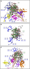
Interaction interfaces of the Ebp1-ribosome complex. (A) Ebp1 (green)-ribosome (60S subunit [rRNA in light blue and ribosomal proteins in gray]) complex is represented as surface in two orientations. (B) Overview of Ebp1 binding site on human ribosome. Ebp1 (green), eL19 (yellow), uL23 (magenta), uL24 (orange), uL29 (wheat) proteins, fragment of 5.8S rRNA (purple), and different regions (H53, H59, and ES27L) of the 28S rRNA (light blue) are shown as cartoons. (CE) Closer view of interaction interface of Ebp1 (green) with eL19 (yellow) and H59 (light blue), uL23 (magenta), and H53 (light blue), and uL29 (wheat) and 5.8S rRNA (purple). Interacting residues are shown as sticks, and polar interactions are marked by dotted lines.
FIGURE 2.
FIGURE 2.
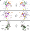
Comparison of the human Ebp1-80S and yeast Arx1-60S complexes (Greber et al. 2016). (A) Superposition of yeast Arx1-ribosome complex (PDB: 5APN [Greber et al. 2016]) on Ebp1-ribosome complex. Ebp1 (green), Arx1 (dark beige), eL19 (yellow), uL23 (magenta), uL24 (orange), uL29 (wheat) proteins, fragment of 5.8S rRNA (purple), and different regions (H53, H59, and ES27L) of the 28S/25S rRNA (light blue and blue) are shown as cartoon. Insert domains of Ebp1 and Arx1 are highlighted with a box. (B) Rotation of ES27L in human Ebp1 (green)-ribosome complex in comparison with yeast Arx1 (dark beige)-60S complex. Only 28S/25S rRNA of human and yeast ribosome are displayed (light blue and blue) for clarity. (C) Additional insertion regions of yeast Arx1 (dark beige) that mediate interaction with uL24 (orange), uL29 (wheat), 5.8S rRNA (purple), and H24 of 28S/25S rRNA are indicated with boxes. Other proteins and rRNA fragments are colored as in A.
FIGURE 3.
FIGURE 3.
Ribosome bound Ebp1 can undergo a rotation around the insert domain. (A) Overview of Ebp1 binding site on human ribosome obtained in this study is shown in two orientations. Ebp1 (green), eL19 (yellow), uL23 (magenta), uL24 (orange), uL29 (wheat) proteins, fragment of 5.8S rRNA (purple), and different regions (H53, H59, and ES27L) of the 28S rRNA (light blue) are shown as cartoon. (B) Overview of Ebp1 binding site on human ribosome from a previous study is shown in two orientations (PDB: 6SXO [Wild et al. 2020]). Ebp1 (light pink), eL19 (yellow), uL23 (magenta), uL24 (orange), uL29 (wheat) proteins, fragment of 5.8S rRNA (purple), and different regions (H53, H59, and ES27L) of the 28S rRNA (light blue) are shown as cartoon (as in A). (C) Relative orientation of Ebp1 (green and light pink) with respect to 60S subunit is shown based on superposition of ribosomal proteins in two orientations. The axis and degree of rotation of Ebp1 are indicated. Arrows (gray) indicate a few regions of Ebp1 that undergo conformational change.
FIGURE 4.
FIGURE 4.
Rotation of Ebp1 leads to remodeling of its interactions with the ribosome. Two conformations of Ebp1 (green and light pink [PDB: 6SXO; Wild et al. 2020]) bound to the 80S ribosome are shown. Fragments of 28S rRNA are displayed in light blue. Loss of interaction with uL24 (orange) is indicated by an arrow.

References

    1. Adams PD, Afonine PV, Bunkóczi G, Chen VB, Davis IW, Echols N, Headd JJ, Hung L-W, Kapral GJ, Grosse-Kunstleve RW, et al. 2010. PHENIX: a comprehensive Python-based system for macromolecular structure solution. Acta Crystallogr D Biol Crystallogr 66: 213–221. 10.1107/S0907444909052925 - DOI - PMC - PubMed
    1. Afonine PV, Klaholz BP, Moriarty NW, Poon BK, Sobolev OV, Terwilliger TC, Adams PD, Urzhumtsev A. 2018a. New tools for the analysis and validation of cryo-EM maps and atomic models. Acta Crystallogr D Struct Biol 74: 814–840. 10.1107/S2059798318009324 - DOI - PMC - PubMed
    1. Afonine PV, Poon BK, Read RJ, Sobolev OV, Terwilliger TC, Urzhumtsev A, Adams PD. 2018b. Real-space refinement in PHENIX for cryo-EM and crystallography. Acta Crystallogr D Struct Biol 74: 531–544. 10.1107/S2059798318006551 - DOI - PMC - PubMed
    1. Anger AM, Armache J-P, Berninghausen O, Habeck M, Subklewe M, Wilson DN, Beckmann R. 2013. Structures of the human and Drosophila 80S ribosome. Nature 497: 80–85. 10.1038/nature12104 - DOI - PubMed
    1. Ball LA, Kaesberg P. 1973. Cleavage of the N-terminal formymlethionine residue from a bacteriophage coat protein in vitro. J Mol Biol 79: 531–537. 10.1016/0022-2836(73)90404-X - DOI - PubMed

Publication types

MeSH terms

LinkOut - more resources