. 2021 Feb 9;118(6):e2017715118.
doi: 10.1073/pnas.2017715118.

Dynamic competition between SARS-CoV-2 NSP1 and mRNA on the human ribosome inhibits translation initiation

Affiliations

Dynamic competition between SARS-CoV-2 NSP1 and mRNA on the human ribosome inhibits translation initiation

Christopher P Lapointe et al. Proc Natl Acad Sci U S A. .

Abstract

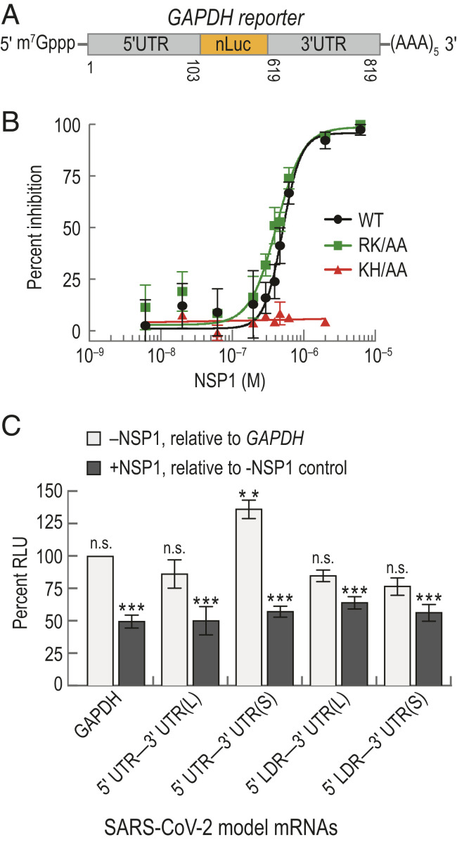
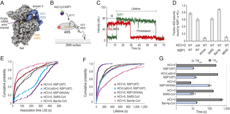
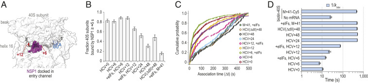
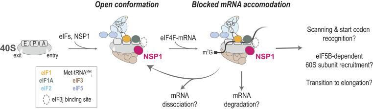
Severe acute respiratory syndrome coronavirus 2 (SARS-CoV-2) is a beta-CoV that recently emerged as a human pathogen and is the causative agent of the COVID-19 pandemic. A molecular framework of how the virus manipulates host cellular machinery to facilitate infection remains unclear. Here, we focus on SARS-CoV-2 NSP1, which is proposed to be a virulence factor that inhibits protein synthesis by directly binding the human ribosome. We demonstrate biochemically that NSP1 inhibits translation of model human and SARS-CoV-2 messenger RNAs (mRNAs). NSP1 specifically binds to the small (40S) ribosomal subunit, which is required for translation inhibition. Using single-molecule fluorescence assays to monitor NSP1-40S subunit binding in real time, we determine that eukaryotic translation initiation factors (eIFs) allosterically modulate the interaction of NSP1 with ribosomal preinitiation complexes in the absence of mRNA. We further elucidate that NSP1 competes with RNA segments downstream of the start codon to bind the 40S subunit and that the protein is unable to associate rapidly with 80S ribosomes assembled on an mRNA. Collectively, our findings support a model where NSP1 proteins from viruses in at least two subgenera of beta-CoVs associate with the open head conformation of the 40S subunit to inhibit an early step of translation, by preventing accommodation of mRNA within the entry channel.

Keywords: NSP1; SARS-CoV-2; eukaryotic translation initiation; human ribosome; single-molecule fluorescence.


Conflict of interest statement

The authors declare no competing interest.

Figures

Fig. 1.
Fig. 1.
NSP1 from SARS-CoV-2 inhibited translation. (A) Schematic of the host model mRNA used in HeLa cell-free IVT assays. The nLuc coding sequence of the mRNA was flanked by the 5′ and 3′ UTRs of human GAPDH. Numbers refer to the nucleotide position in NCBI GenBank accession: AF261085. (B) NSP1 dose–response analysis of GAPDH reporter mRNA IVT in HeLa extract treated with either wild-type (WT; n = 3), a predicted ribosome-binding−deficient mutant (KH/AA; n = 2), or an RNA cleavage-deficient mutant (RK/AA; n = 2) NSP1. The mean response ± SEM (symbols, error bars) and curve fits (lines) from nonlinear regression analysis of the data are plotted. WT IC50 = 510 ± 20 nM (95% CI; R2 = 0. 83), and RK/AA IC50 = 420 ± 11 nM (95% CI; R2 = 0.89). (C) Plot of the mean nLuc relative light unit (RLU) signal from cell-free translation of host and viral reporter mRNAs in the absence and presence (400 nM) of wild-type NSP1. Without NSP1 (light gray), mean translational activities (percent RLU) were compared to GAPDH reporter mRNA in the absence of NSP1 (** = P ≤ 0.0006; and n.s. = P ≥ 0.2, one-way ANOVA). GAPDH (n = 6), 5′ UTR−3′ UTR(S) mRNA (n = 3), 5′ UTR−3′ UTR(L) (n = 3), and 5′ LDR reporter mRNAs (n = 5). In the presence of NSP1 (dark gray), samples were compared to a control reaction that lacked NSP1 (p0.0008, t test). GAPDH (n = 6), viral 5′ UTR mRNAs (n = 3), and viral 5′ LDR mRNAs (n = 2). Error bars represent SEM.
Fig. 2.
Fig. 2.
NSP1 associated with 40S subunits and most ribosomal preinitiation complexes. (A) Experimental setup. Using a ZMW system, 40S ribosomal subunits biotinylated on RACK1 were tethered to a neutravidin-coated imaging surface within thousands of individual ZMWs (also see SI Appendix, Fig. S3B). Upon start of data acquisition, Cy3-NSP1 (N-terminal ybbR tag) was added, and fluorescence intensities were monitored. (B) Example single-molecule fluorescence trace that depicts association of Cy3-NSP1 with a tethered eIF1–40S subunit complex. Prior to tethering, 40S subunits were incubated with 30-fold molar excess eIF1. During imaging, eIF1 was present at 1 µM. The association time (Δt) was defined as the time elapsed from the addition of Cy3-NSP1 until the burst of Cy3 fluorescence (green), which signified NSP1 association. The lifetime was defined as the duration of the Cy3 fluorescence signal. (C) Plot of the fraction of ZMWs that contained at least one Cy3-NSP1 binding event ≥∼5 s in duration in the indicated conditions at 20 °C. Error bars represent 99% CI. (D) Plot of apparent association rates (open circles) of Cy3-NSP1 with tethered eIF1–40S subunit complexes at the indicated NSP1 concentrations at 20 °C. The dashed line represents a fit from linear regression analysis (adjusted R2 = 0.99), with a slope of 0.3 ± 0.1 and y intercept of 0.0013 ± 0.002 (errors represent 95% CI). Error bars on the open circles represent 95% CI of the rates. (E) Plot of the cumulative probability of Cy3-NSP1 association times with the indicated ribosomal preinitiation complexes. Cy3-NSP1 was present at 25 nM (final concentration), and the temperature was 30 °C. The eIFs were preincubated with 40S subunits, and they were included at molar excess relative to 40S subunits during tethering and imaging to promote formation of the indicated complexes. The proteins eIF1, eIF1A, and eIF5 were present at 1 µM; the eIF2-GMPPNP-Met-tRNAiMet ternary complex at 100 nM; and eIF3Δj at 50 nM. Lines represent fits to double-exponential functions. See SI Appendix, Table S2 for samples sizes and the parameters for relevant fits. (F) Plot of Cy3-NSP1 median association times (light blue) with the indicated ribosomal preinitiation complexes. Error bars represent 95% CI of the median values.
Fig. 3.
Fig. 3.
NSP1 preferentially associated with the open head conformation of the 40S subunit. (A) Model of the human 40S subunit (gray) bound by the HCV IRES (blue) (PDB ID code 5A2Q) (38). This model of the IRES ends at the start codon (AUG, highlighted in orange), leaving the mRNA entry channel of the 40S subunit empty. Domain II of the IRES holds the head of the 40S subunit in the open conformation. (B) Schematic of the single-molecule fluorescence assay. The 40S ribosomal subunits were labeled with Cy5 dye via RACK1-ybbR. Preformed IRES–40S-Cy5 complexes were tethered to the ZMW imaging surface. At the start of data acquisition, Cy3-NSP1 (N-terminal ybbR tag) was added at 25 nM (final concentration) at 30 °C. (C) Example single-molecule fluorescence trace that depicts a tethered 40S–HCV+0 complex and subsequent association of NSP1. The 40S subunit and ybbR-NSP1 were labeled with Cy5 (red) and Cy3 (green) dyes, respectively. Loss of fluorescence signal due to dye photobleaching is indicated. Raw fluorescence intensities were corrected in this image to set baseline intensities to zero for presentation. The association time (Δt) was defined as time elapsed from the addition of Cy3-NSP1 until the burst of Cy3 fluorescence (green), which signified NSP1 association. The lifetime was defined as the duration of the Cy3 fluorescence signal. (D) Plot of the fraction of the indicated IRES–40S subunit complexes bound at least once by the indicated NSP1 protein for ≥∼5 s. Error bars represent 99% CI. WT, SARS-CoV-2 NSP1; KH/AA, SARS-CoV-2 NSP1(KH/AA). (E and F) Plot of the cumulative probability of observed Cy3-NSP1 association times (E) and lifetimes (F) with the indicated IRES–40S subunit complexes at 30 °C. The indicated Cy3-NSP1 was added at 25 nM (final concentration) in all experiments. Lines represent fits to double-exponential functions. See SI Appendix, Table S3 for samples sizes and the parameters for relevant fits. Association times were determined with the excitation laser (532 nm) at 0.6 µW/µm2, whereas lifetimes were determined at the further reduced power of 0.1 µW/µm2 to enhance dye stability. (G) Plot of the reciprocal apparent association (kobs) (light blue) and dissociation (koff) (light gray) rates of the indicated NSP1 binding to the indicated IRES–40S subunit complexes. Rates were derived from fits of data to double-exponential functions, with the fast association rate and predominate lifetime reported here. See SI Appendix, Table S3 for samples sizes and all parameters from relevant fits. Error bars represent 95% CI.
Fig. 4.
Fig. 4.
The mRNA within the entry channel of the 40S subunit inhibited SARS-CoV-2 NSP1 association. (A) Image of the intersubunit interface of the human 40S ribosomal subunit, with the approximate positions of mRNA (gray) and NSP1 (purple) modeled to show the predicted steric clash when segments of RNA longer than 6 nt are downstream of the start codon (blue). Models were aligned using ChimeraX (mmaker command) and PDB ID codes 6ZLW (19) and 6YAL (76). (B) Plot of the fraction of the indicated mRNA–40S subunit complexes bound at least once by SARS-CoV-2 NSP1 for ≥∼5 s. Error bars represent 99% CI. (C) Plot of the cumulative probability of observed Cy3-NSP1 association times with the indicated mRNA–40S indicates eIF1, eIF1A, eIF3Δj, eIF5, and TC(GMPPNP) were included at all stages of the experiment. Lines represent fits to single- or double-exponential functions. See SI Appendix, Table S4 for samples sizes and the parameters for relevant fits. (D) Plot of the reciprocal apparent association rates (kobs) (light blue) of SARS-CoV-2 NSP1 binding to the indicated mRNA–40S subunit complexes, derived from fits of the data to double-exponential functions, with the fast association rate reported here. See SI Appendix, Table S4 for samples sizes and the parameters from relevant fits. Error bars represent 95% CI.
Fig. 5.
Fig. 5.
NSP1 inefficiently associated with 80S ribosomes assembled on the CrPV IRES. (A) Model of the human 80S ribosome (PDB ID code 4UG0). The 40S and 60S subunits are in gray and pink, respectively. The ybbR tag was fused to either the N terminus of uS19 (green, 40S) or the C terminus of uL18 (red, 60S). Based on available structural models, the ybbR tags were predicted to be within FRET distance in translation-competent 80S ribosomes. (B) Example fluorescent trace and calculated FRET efficiency plot. The 40S-ybbR-Cy3 and 60S-ybbR-Cy5 subunits were incubated with the CrPV IRES to assemble 80S ribosomes on the biotinylated RNA. Following tethering of the complex, molecules were imaged at equilibrium using TIRFM. Molecules were expected to begin in a Cy3 (green, FRET donor) to Cy5 (red, acceptor) FRET state, followed by photobleaching of both dyes. The region of the trace that corresponds to FRET is highlighted by the gray box. (C) Plot of the distribution of observed FRET efficiencies for the intersubunit FRET signal on 80S ribosomes. Frequencies of observed FRET efficiencies were binned into 35 bins (open circles) across the indicated range. The line represents a fit to a single-Gaussian function, which yielded a mean FRET efficiency of 0.5 ± 0.01 (95% CI); n = 104. (D) Schematic of the single-molecule fluorescence assay. The 40S ribosomal subunits were labeled with Cy3 dye via uS19-ybbR, and 60S subunits were labeled with Cy5 via uL18-ybbR. Preformed 80S–CrPV IRES complexes were tethered to the ZMW imaging surface. At the start of data acquisition, Cy5.5-NSP1 (N-terminal ybbR tag) was added at 25 nM (final concentration) at 30 °C. (E) Example single-molecule fluorescence traces that depict addition of Cy5.5-NSP1 to tethered CrPV+1 RNAs bound by 80S ribosomes. The 40S subunit was labeled with Cy3 (green), 60S subunit with Cy5 (red), and NSP1 with Cy5.5 (magenta). The two traces are from the same experiment where 80S–CrPV+1 complexes were tethered. Top trace depicts a complex with an NSP1 binding event (∼30% of traces), and Bottom trace lacks an NSP1 event (∼70% of traces). Raw fluorescence intensities were corrected in this image to set baseline intensities to zero for presentation. Due to bleed through across the three fluorescent channels, the Cy3, Cy5, and Cy5.5 signals were made transparent before and after relevant events for presentation here. The association time (Δt) was defined as the time elapsed from the addition of Cy5.5-NSP1 until the burst of Cy5.5 fluorescence (magenta), which signified NSP1 association. The lifetime was defined as the duration of the Cy5.5 fluorescence signal. (F) Plot of the cumulative probability of observed NSP1 association times with the indicated ribosomal–CrPV IRES complexes at 30 °C. Cy5.5-NSP1 was added at 25 nM (final concentration). Lines represent fits to double-exponential functions. See SI Appendix, Table S5 for samples sizes and the parameters for relevant fits.
Fig. 6.
Fig. 6.
NSP1 remained bound to 40S subunits upon association with model mRNAs. (A) Plot of the fraction of stable (5s) 40S subunit binding events that coassociated with NSP1, as defined in B. Error bars represent 99% CI. (B) Example single-molecule fluorescence trace that depicts association of NSP1–40S subunit complexes with a tethered HCV+0 IRES molecule. The 40S subunit and NSP1 were labeled with Cy3 (green) and Cy5.5 (magenta) dyes, respectively. Raw fluorescence intensities were corrected in this image to set baseline intensities to zero for presentation. The initial NSP1–40S subunit association time (Δt1) was defined as the time elapsed from the addition of the complex until the burst of Cy3 and Cy5.5 fluorescence, which signified association of the NSP1-40S subunit complex with the tethered IRES. In experiments that lacked NSP1, Δt1 was defined using the first burst of Cy3 signal alone. The 40S subunit lifetime (τ1) was defined as the duration of the Cy3 fluorescence signal. The initial NSP1 lifetime (NSP1τ1) was defined as the duration of the Cy5.5 signal that coappeared with the Cy3 signal. For NSP1 reassociation analyses, we focused on ZMWs where a single 40S subunit associated within the first 200 s (∼75% of all events; see SI Appendix, Fig. S8F). We then quantified the time elapsed from the loss of the first Cy5.5 signal to the next burst of Cy5.5 fluorescence at least ∼20 s in length (∼70% of initial NSP1 binding events), which was defined as the NSP1 reassociation time (NSP1 Δt2). The duration of this second Cy5.5 event was defined as the reassociated NSP1 lifetime (NSP1τ2). (C and D) Plots of the indicated lifetimes. Here, all experiments were conducted in the presence of NSP1. See SI Appendix, Fig. S8 D and E for 40S subunit lifetimes in the absence of NSP1. Lifetimes were defined as the reciprocal of the predominate dissociation rate derived from fits to double-exponential functions, with error bars representing the 95% CI of the rate. See SI Appendix, Table S6 for samples sizes and the parameters for relevant fits. (E) Time-of-addition cell-free IVT experimental design. GAPDH reporter mRNA and WT NSP1 (400 nM) were added to HeLa IVT reactions in the order depicted above. The nLuc signal was continuously monitored in situ. Using the same color scheme as E, FI depict results from six independent replicates for each experimental condition, except for the “preincubation with NSP1 reaction” (red bar in E), which has n = 3. (F) Time course of nLuc synthesis from a representative time-of-addition experiment. (G) Representative Gaussian fits of the second derivative of nLuc synthesis time course data shown in F. (H) Plot of mean synthesis time. Error bars represent SEM, *P < 0.0001. (I) Plot of translational productivity. Error bars represent SEM, *P = 0.045, one-way ANOVA.
Fig. 7.
Fig. 7.
Proposed model. In the absence of eIF3j, NSP1 preferentially associates with the open conformation of the 40S subunit to block full accommodation of the mRNA in the entry channel, which inhibits translation initiation. How incomplete mRNA accommodation impacts mRNA recruitment, scanning, start codon selection, 60S subunit recruitment, and the transition to elongation remain open questions. Whether and how NSP1–40S subunit complexes lead to mRNA degradation is also unknown.

Similar articles

Cited by

References

    1. Cui J., Li F., Shi Z.-L., Origin and evolution of pathogenic coronaviruses. Nat. Rev. Microbiol. 17, 181–192 (2019). - PMC - PubMed
    1. Drosten C., et al. ., Identification of a novel coronavirus in patients with severe acute respiratory syndrome. N. Engl. J. Med. 348, 1967–1976 (2003). - PubMed
    1. Ksiazek T. G.et al. .; SARS Working Group , A novel coronavirus associated with severe acute respiratory syndrome. N. Engl. J. Med. 348, 1953–1966 (2003). - PubMed
    1. Zaki A. M., van Boheemen S., Bestebroer T. M., Osterhaus A. D. M. E., Fouchier R. A. M., Isolation of a novel coronavirus from a man with pneumonia in Saudi Arabia. N. Engl. J. Med. 367, 1814–1820 (2012). - PubMed
    1. Zhou P., et al. ., A pneumonia outbreak associated with a new coronavirus of probable bat origin. Nature 579, 270–273 (2020). - PMC - PubMed

Publication types

MeSH terms