. 2021 Jan 21;12(1):491.
doi: 10.1038/s41467-020-20680-5.

White pupae phenotype of tephritids is caused by parallel mutations of a MFS transporter

Affiliations

White pupae phenotype of tephritids is caused by parallel mutations of a MFS transporter

Christopher M Ward et al. Nat Commun. .

Abstract

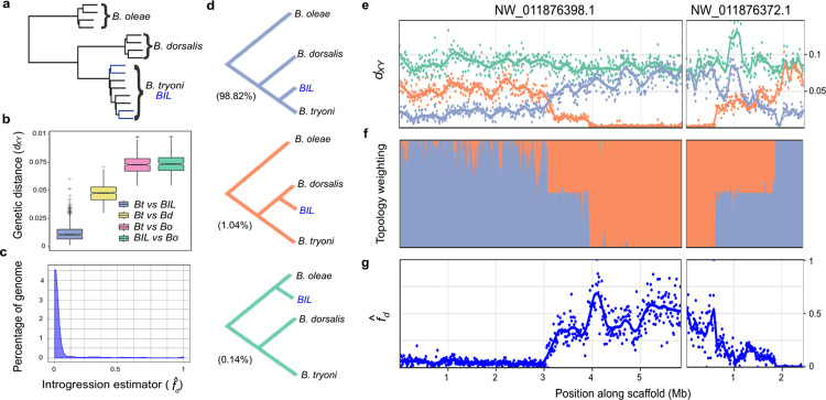
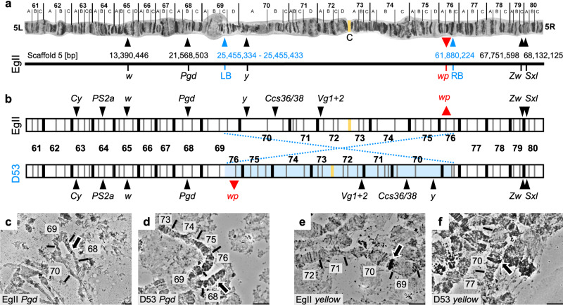
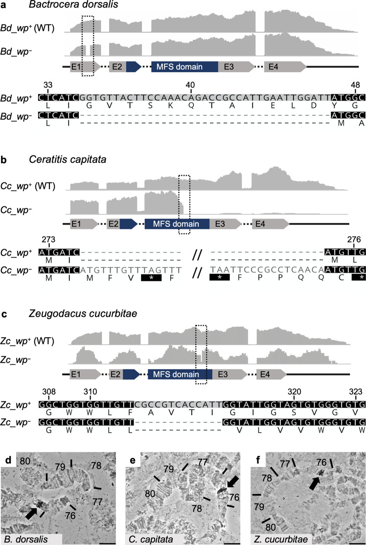
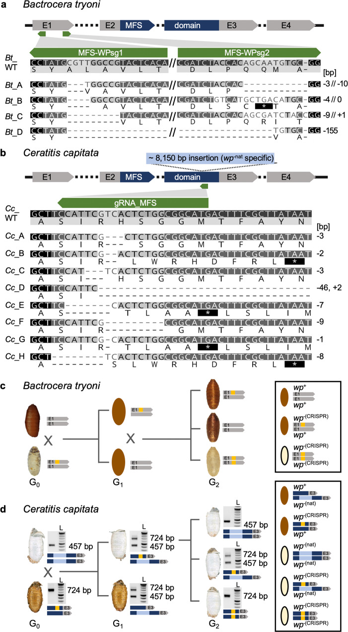
Mass releases of sterilized male insects, in the frame of sterile insect technique programs, have helped suppress insect pest populations since the 1950s. In the major horticultural pests Bactrocera dorsalis, Ceratitis capitata, and Zeugodacus cucurbitae, a key phenotype white pupae (wp) has been used for decades to selectively remove females before releases, yet the gene responsible remained unknown. Here, we use classical and modern genetic approaches to identify and functionally characterize causal wp- mutations in these distantly related fruit fly species. We find that the wp phenotype is produced by parallel mutations in a single, conserved gene. CRISPR/Cas9-mediated knockout of the wp gene leads to the rapid generation of white pupae strains in C. capitata and B. tryoni. The conserved phenotype and independent nature of wp- mutations suggest this technique can provide a generic approach to produce sexing strains in other major medical and agricultural insect pests.


Conflict of interest statement

The authors declare no competing interests.

Figures

Fig. 1
Fig. 1. Characterization of total introgression from B. dorsalis into the Bactrocera introgressed line and identification of the white pupae locus.
a Species tree constructed from 1846 single copy ortholog gene trees for four haplotypes of B. oleae, B. dorsalis, B. tryoni, and BIL. Branches corresponding to BIL individuals are shown in blue. All nodes were well supported with posterior probabilities >0.97. b Nei’s absolute genetic distance (dXY) calculated for tiled 100 kb windows across the genome between B. tryoni vs BIL (Bt vs BIL); B. tryoni vs B. dorsalis (Bt vs Bd); B. tryoni vs B. oleae (Bt vs Bo); and BIL vs B. oleae (BIL vs Bo). Box and whisker graphs (including outliers) represent a summary of 2294 genomic windows. Boxes show the first and third inter quartile range (IQR) while whiskers extend to a maximum of 1.5 ∗ IQR. All values outside 1.5 ∗ IQR are shown as plus signs. c The introgression estimator (ƒd) calculated across tiled 100 kb windows to identify regions of disproportionately shared alleles between BIL and B. dorsalis, ƒd (Bt, BIL, Bd; Bo). d The three evolutionary hypothesis/topologies of interest to identify introgressed regions and their representation across the genome: species (purple, 98.82%), introgression (orange, 1.04%) and a negative control tree (green, 0.14%). e Nei’s absolute genetic distance (dXY) calculated for tiled 10 kb windows across the candidate wp locus for B. tryoni vs BIL (purple), B. dorsalis vs BIL (orange), B. oleae vs BIL (green). f Topology weighting for each topology shown in d, calculated for 1 kb tiled local trees across the candidate wp locus. g The introgression estimator (ƒd) calculated across tiled 10 kb windows for the comparison ƒd (Bt, BIL, Bd; Bo) to identify the start and end of the introgressed locus. Source data are provided in a Source Data file.
Fig. 2
Fig. 2. Genomic positioning of the D53 inversion on chromosome 5 of C. capitata.
a Chromosome scale assembly of C. capitata EgII chromosome 5. Shown are the positions of in situ mapped genes white (w), 6-phosphogluconate dehydrogenase (Pgd), glucose-6-phosphate 1-dehydrogenase (Zw), and sex lethal (Sxl), the position of the D53 inversion breakpoints (blue; LB = left breakpoint, RB = right breakpoint), and the relative position of white pupae (wp) on the polytene chromosome map of chromosome 5 (left (L) and right (R) chromosome arm, linked at the centromeric region (C)) and the PacBio-Hi-C EgII scaffold_5 (bp = base pairs), representing the complete chromosome 5 (Ccap3.2.1, accession GCA_905071925). The position of the yellow gene (y, LOC101455502) was confirmed on chromosome 5 70A by in situ hybridization, despite its sequence not been found in the scaffold assembly. b Schematic illustration of chromosome 5 without (EgII, WT) and with (D53) D53 inversion, with additional marker genes Curly (Cy), integrin-aPS2 (PS2a), white (w), chorion S36/38 (Ccs36/38), vitellogenin-1/2-like (Vg1 + 2). The inverted part of chromosome 5 is shown in light blue, the centromere in yellow. Two probes, one inside (y, 70A) and one outside (Pgd, 68B) of the left inversion breakpoint were used to verify the D53 inversion breakpoints by in situ hybridization. WT EgII is shown in c and e, D53 in d and f. Chromosomal segments are numbered, arrows in micrographs indicate in situ hybridization signal. In situ hybridizations were done at least in duplicates and at least ten nuclei were analyzed per sample, scale bar = 10 µm. All replicates led to similar results. The source data underlying Fig. 2c–f are provided as a Source Data file.
Fig. 3
Fig. 3. Identification of the wp mutation in the transcriptomes of B. dorsalis, C. capitata, and Z. cucurbitae.
The gray graphs show expression profiles from the candidate wp loci in WT (wp+) and mutant (wp) flies at the immobile pupae stages of a B. dorsalis, b C. capitata, and c Z. cucurbitae. The gene structure (not drawn to scale) is indicated below as exons (arrows labeled E1–E4) and introns (dashed lines), the Major Facilitator Superfamily (MFS) domain is shown in blue. The positions of independent wp mutations (Bd: 37 bp deletion, Cc: approximate 8150 bp insertion, Zc: 13 bp deletion) are marked with black dashed boxes in the expression profiles and are shown in detail below the gene models based on de novo assembly of RNAseq data from WT and white pupae phenotype individuals (nucleotide and amino acid sequences). Deletions are shown as dashes, alterations on protein level leading to premature stop codons are depicted as asterisks highlighted in black. In situ hybridization on polytene chromosomes for d B. dorsalis, e C. capitata, and f Z. cucurbitae confirmed the presence of the wp locus on the right arm of chromosome 5 in all three species (arrows in micrographs). In situ hybridizations were done at least in duplicates and at least ten nuclei were analyzed per sample, scale bar = 10 µm. The source data underlying Fig. 3d–f are provided as a Source Data file.
Fig. 4
Fig. 4. CRISPR/Cas9-based generation of homozygous wp-(CRISPR) lines in B. tryoni and C. capitata.
A schematic structure of the wp CDS exons (E1, E2, E3, E4) including the MFS domain in B. tryoni (a) and C. capitata (b) are shown. Positions of gRNAs targeting the first and third exon in B. tryoni and C. capitata, respectively, are indicated by green arrows. Nucleotide and amino acid sequences of mutant wp alleles identified in G1 individuals are compared to the WT reference sequence in B. tryoni (a) and C. capitata (b). Deletions are shown as dashes, alterations on protein level leading to premature stop codons are depicted as asterisks highlighted in black. Numbers on the right side represent InDel sizes (bp = base pairs). Crossing schemes to generate homozygous wp−(CRISPR) lines in B. tryoni (c) and C. capitata (d) show different strategies to generate wp strains. Bright-field images of empty puparia are depicted for both species. Genotype schematics and corresponding PCR analysis (for C. capitata) validating the presence of CRISPR-induced (orange) and natural (blue, for C. capitata) wp mutations are shown next to the images of the puparia. c Injected G0 B. tryoni were backcrossed to the Ourimbah laboratory strain resulting in uniformly brown G1 offspring (depicted as illustration because no images were acquired during G1). G1 inbreeding led to G2 individuals homozygous for the white pupae phenotype. d Injected WT G0 flies were crossed to flies homozygous for the naturally occurring wp allele (wp−(nat)). wp−(nat) (457 bp amplicon) and wp−(CRISPR) or WT (724 bp amplicon) alleles were identified by multiplex PCR (left lane; L = NEB 2 log ladder). White pupae phenotypes in G1 indicated positive CRISPR events. G2 flies with a white pupae phenotype that were homozygous for the wp−(CRISPR) allele were used to establish lines. PCR was done once for each individual, wp−(CRISPR) alleles were verified and further analyzed via sequencing. The source data underlying Fig. 4d are provided as a Source Data file.

Similar articles

Cited by

References

    1. Robinson, A. S. & Hooper, G. Fruit Flies: Their Biology, Natural Enemies, and Control Vol. 1 (Elsevier, 1989).
    1. Suckling DM, et al. Eradication of tephritid fruit fly pest populations: outcomes and prospects. Pest Manag. Sci. 2016;72:456–465. doi: 10.1002/ps.3905. - DOI - PubMed
    1. Dyck, V. A. et al. Sterile Insect Technique – Principles and Practice in Area-Wide Integrated Pest Management (eds Dyck, V. A., Hendrichs, J. & Robinson, A. S.) (Springer, 2005).
    1. Vreysen, M., Robinson, A. S. & Hendrichs, J. Area-Wide Control of Insect Pests: from Research to Field Implementation (Springer, 2007).
    1. Rendon P, McInnis D, Lance D, Stewart J. Medfly (Diptera: Tephritidae) genetic sexing: large-scale field comparison of males-only and bisexual sterile fly releases in Guatemala. J. Econ. Entomol. 2004;97:1547–1553. doi: 10.1603/0022-0493-97.5.1547. - DOI - PubMed

Publication types

Substances

LinkOut - more resources