. 2021 Jan 21;12(1):498.
doi: 10.1038/s41467-020-20603-4.

Sarcoma classification by DNA methylation profiling

Christian Koelsche #  1   2   3 Daniel Schrimpf #  1   2 Damian Stichel #  2 Martin Sill #  4   5 Felix Sahm  1   2 David E Reuss  1   2 Mirjam Blattner  4   6 Barbara Worst  4   6   7 Christoph E Heilig  8 Katja Beck  8   9 Peter Horak  8 Simon Kreutzfeldt  8 Elke Paff  4   6   7 Sebastian Stark  4   6   7 Pascal Johann  4   6   7 Florian Selt  4   7   10 Jonas Ecker  4   7   10 Dominik Sturm  4   6   7 Kristian W Pajtler  4   5   7 Annekathrin Reinhardt  1   2 Annika K Wefers  1   2 Philipp Sievers  1   2 Azadeh Ebrahimi  2 Abigail Suwala  1   2 Francisco Fernández-Klett  1   2 Belén Casalini  2 Andrey Korshunov  1   2 Volker Hovestadt  11   12 Felix K F Kommoss  3 Mark Kriegsmann  3 Matthias Schick  13 Melanie Bewerunge-Hudler  13 Till Milde  4   7   10 Olaf Witt  4   7   10 Andreas E Kulozik  4   7 Marcel Kool  4   5 Laura Romero-Pérez  14 Thomas G P Grünewald  14 Thomas Kirchner  15 Wolfgang Wick  16   17 Michael Platten  18   19 Andreas Unterberg  20 Matthias Uhl  21   22 Amir Abdollahi  21   22   23   24 Jürgen Debus  21   22   23   24 Burkhard Lehner  25 Christian Thomas  26 Martin Hasselblatt  26 Werner Paulus  26 Christian Hartmann  27 Ori Staszewski  28   29 Marco Prinz  28   30   31 Jürgen Hench  32 Stephan Frank  32 Yvonne M H Versleijen-Jonkers  33 Marije E Weidema  33 Thomas Mentzel  34 Klaus Griewank  35 Enrique de Álava  36   37 Juan Díaz Martín  36 Miguel A Idoate Gastearena  38 Kenneth Tou-En Chang  39 Sharon Yin Yee Low  40 Adrian Cuevas-Bourdier  41 Michel Mittelbronn  41   42   43   44 Martin Mynarek  45 Stefan Rutkowski  45 Ulrich Schüller  45   46   47 Viktor F Mautner  48 Jens Schittenhelm  49 Jonathan Serrano  50 Matija Snuderl  50 Reinhard Büttner  51 Thomas Klingebiel  52 Rolf Buslei  53 Manfred Gessler  54 Pieter Wesseling  55   56 Winand N M Dinjens  57 Sebastian Brandner  58   59 Zane Jaunmuktane  59   60 Iben Lyskjær  61 Peter Schirmacher  3 Albrecht Stenzinger  3 Benedikt Brors  62 Hanno Glimm  63   64   65   66 Christoph Heining  64   65   66 Oscar M Tirado  67 Miguel Sáinz-Jaspeado  67 Jaume Mora  68 Javier Alonso  69 Xavier Garcia Del Muro  70 Sebastian Moran  71 Manel Esteller  72   73   74   75 Jamal K Benhamida  76 Marc Ladanyi  76 Eva Wardelmann  77 Cristina Antonescu  76 Adrienne Flanagan  78   79 Uta Dirksen  80   81 Peter Hohenberger  82 Daniel Baumhoer  83 Wolfgang Hartmann  84 Christian Vokuhl  85 Uta Flucke  86 Iver Petersen  87   88 Gunhild Mechtersheimer  3 David Capper  89 David T W Jones  4   6 Stefan Fröhling  8 Stefan M Pfister  4   5   7 Andreas von Deimling  90   91
Affiliations

Sarcoma classification by DNA methylation profiling

Christian Koelsche et al. Nat Commun. .

Abstract

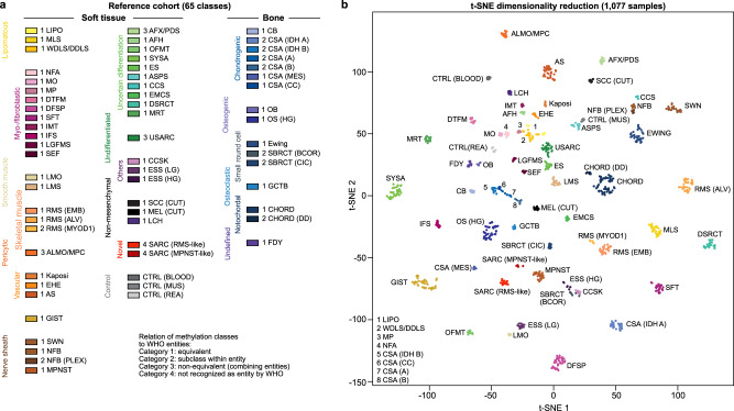
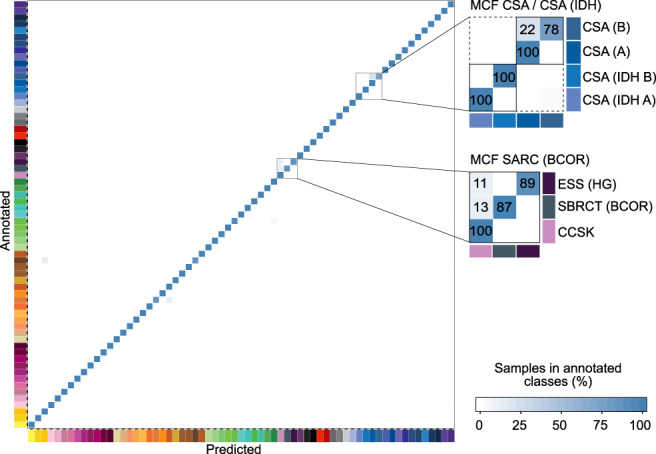
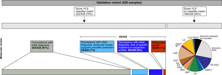
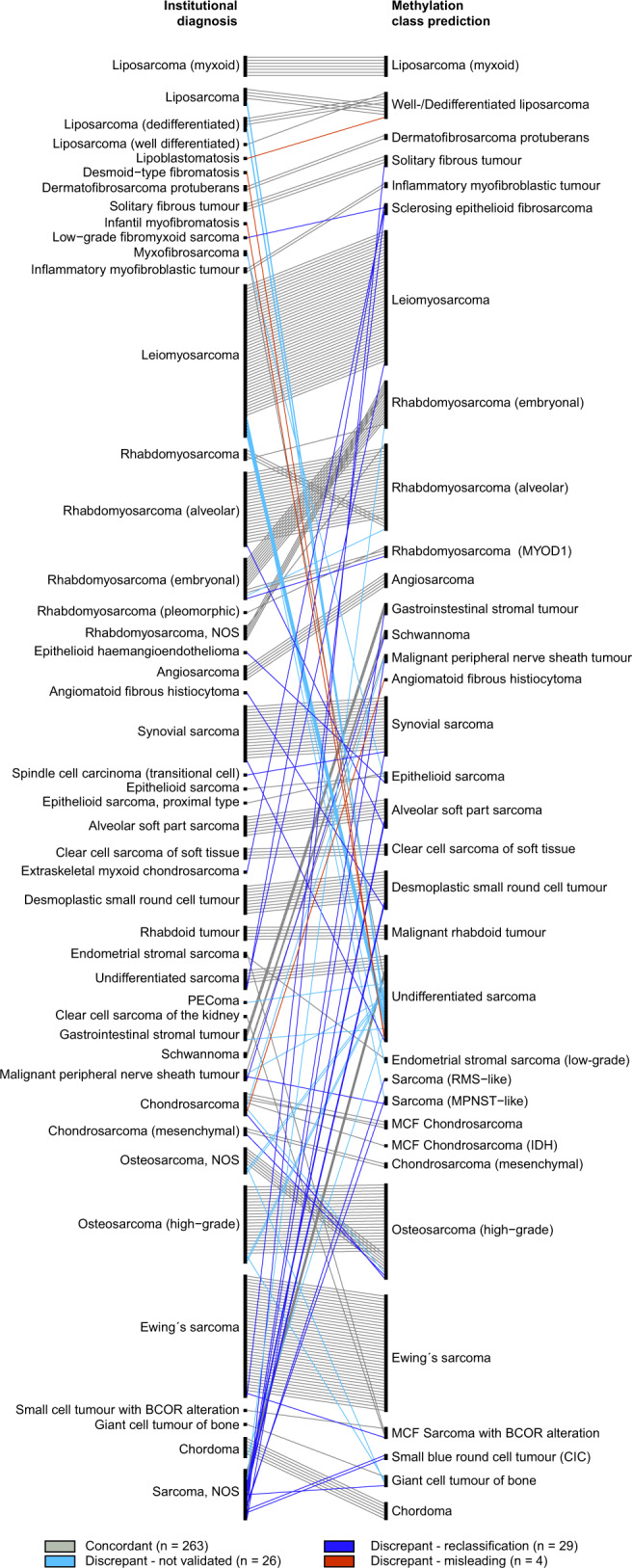
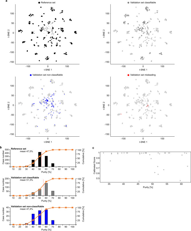
Sarcomas are malignant soft tissue and bone tumours affecting adults, adolescents and children. They represent a morphologically heterogeneous class of tumours and some entities lack defining histopathological features. Therefore, the diagnosis of sarcomas is burdened with a high inter-observer variability and misclassification rate. Here, we demonstrate classification of soft tissue and bone tumours using a machine learning classifier algorithm based on array-generated DNA methylation data. This sarcoma classifier is trained using a dataset of 1077 methylation profiles from comprehensively pre-characterized cases comprising 62 tumour methylation classes constituting a broad range of soft tissue and bone sarcoma subtypes across the entire age spectrum. The performance is validated in a cohort of 428 sarcomatous tumours, of which 322 cases were classified by the sarcoma classifier. Our results demonstrate the potential of the DNA methylation-based sarcoma classification for research and future diagnostic applications.


Conflict of interest statement

A patent for a DNA methylation-based method for classifying tumour species of the brain has been applied for by the Deutsches Krebsforschungszentrum Stiftung des öffentlichen Rechts and Ruprecht-Karls-Universität Heidelberg (EP 3067432 A1) with S.M.P., A.v.D., D.T.W.J., D.C., V.Ho., M.Si., M.B.H. and M.Sc. as inventors. The other authors declare no competing interests.

Figures

Fig. 1
Fig. 1. Establishing the DNA methylation-based sarcoma reference cohort.
a Overview of 62 tumour and three control DNA methylation classes included in the sarcoma classifier reference cohort. The methylation classes are colour-coded and grouped according to the WHO scheme. The relation between methylation classes and the WHO defined subtypes is categorised in 4 tiers: equivalent to a WHO entity (category 1); subgroup of a WHO entity (category 2); combining WHO entities (category 3); non-defined by WHO (category 4). b Visualisation of the reference cohort methylation profiles (n = 1,077) using t-distributed stochastic neighbour embedding (t-SNE) dimensionality reduction. Individual samples are colour-coded in the respective class colour (n = 65) as given in (a). Abbreviations: LIPO, lipoma; MLS, myxoid liposarcoma; WDLS/DDLS, well differentiated liposarcoma/dedifferentiated liposarcoma; NFA, nodular fasciitis; MO, myositis ossificans; MP, myositis proliferans; DTFM, desmoid-type fibromatosis; DFSP, dermatofibrosarcoma protuberans; SFT, solitary fibrous tumour; IMT, inflammatory myofibroblastic tumour; IFS, infantile fibrosarcoma; LGFMS, low-grade fibromyxoid sarcoma; SEF, sclerosing epithelioid fibrosarcoma; LMO, leiomyoma; LMS, leiomyosarcoma; RMS (EMB), embryonal rhabdomyosarcoma; RMS (ALV), alveolar rhabdomyosarcoma; RMS (MYOD1); rhabdomyosarcoma with MYOD1 mutation; ALMO/MPC, angioleiomyoma/myopericytoma; EHE, epithelioid haemangioendothelioma; AS, angiosarcoma; GIST, gastrointestinal stromal tumour; SWN, schwannoma; NFB, neurofibroma; NFB (PLEX), plexiform neurofibroma; MPNST, malignant peripheral nerve sheath tumour; AFX/PDS, atypical fibroxanthoma/pleomorphic dermal sarcoma; AFH, angiomatoid fibrous histiocytoma; OFMT, ossifying fibromyxoid tumour; SYSA, synovial sarcoma; ES, epithelioid sarcoma; ASPS, alveolar soft part sarcoma; CCS, clear cell sarcoma of soft parts; EMCS, extraskeletal myxoid chondrosarcoma; DSRCT, desmoplastic small round cell tumour; MRT, malignant rhabdoid tumour; USARC, undifferentiated sarcoma; CCSK, clear cell sarcoma of the kidney; ESS (LG), low-grade endometrial stromal sarcoma; ESS (HG), high-grade endometrial stromal sarcoma; SCC (CUT), cutaneous squamous cell carcinoma; MEL (CUT), cutaneous melanoma; SARC, sarcoma; CTRL, control; MUS, muscle tissue; REA, reactive tissue; CB, chondroblastoma; CSA, chondrosarcoma; CSA (MES), mesenchymal chondrosarcoma; CSA (CC), clear cell chondrosarcoma; OB, osteoblastoma; OS (HG), high-grade conventional osteosarcoma; SBRCT, small blue round cell tumour; GCTB, giant cell tumour of bone; CHORD, chordoma; DD, dedifferentiated; FDY, fibrous dysplasia; LCH, Langerhans cell histiocytosis.
Fig. 2
Fig. 2. Cross-validation of the DNA methylation-based sarcoma classifier.
Heat map showing results of a threefold cross-validation of the Random Forest classifier incorporating information of n = 1077 biologically independent samples allocated to 65 methylation classes. Deviations from the bisecting line represent misclassification errors (using the maximum calibrated score for class prediction). Methylation class families (MCF) are indicated by black squares. The colour code and abbreviations are identical to Fig. 1a. Numbers of this figure are summarized in Supplementary Data 4.
Fig. 3
Fig. 3. Validation of the sarcoma classifier.
In total, 426 independent sarcoma samples were analysed. 75% matched to an established DNA methylation class with a classifier prediction cut-off score of ≥0.9. 25% reached a classifier prediction cut-off score of <0.9. Abbreviations are identical to Fig. 1a.
Fig. 4
Fig. 4. Comparison of pathological diagnosis and methylation class prediction.
Classifier validation using sarcoma cases enrolled in the MNP2.0, PTT2.0, INFORM or NCT MASTER trials. Institutional diagnosis (left) and classifier prediction (right) of the 322 cases that received a methylation class prediction ≥0.9. The institutional diagnosis of 263 cases matched the classifier prediction (concordant; grey bars). In 59 cases the classifier prediction differed from institutional diagnosis, with 29 cases reclassified in favour of the methylation class prediction (discrepant—reclassified; blue bars), 26 cases where molecular validation analysis was inconclusive (discrepant; light blue bars), and four cases with a misleading classifier result (discrepant – misleading; red bar).
Fig. 5
Fig. 5. Impact of tumour cell purity on classifier performance.
a Unsupervised clustering of the combined reference (n = 1077) and diagnostic cohort (n = 428) using t-SNE dimensionality reduction. The reference set is indicated in the upper left plot. The diagnostic samples coded as classifiable (n = 318, grey dots; upper right plot), non-classifiable (n = 106, blue dots; lower left plot) and misleading (n = 4, red dots; lower right plot). The classifiable cases show high overlap with the reference cases. The non-classifiable cases frequently fall in the periphery of or are completely separate from the reference samples. b Tumour cell purity histogram plots of the reference set and the validation set subdivided into classifiable and non-classifiable cases. The mean value is indicated as dashed red line and provided as number [%]. c Tumour cell purity plotted against calibrated score for conventional osteosarcoma cases of the validation set.

References

    1. Fletcher, C. D. M., Bridge, J. A., Hogendoorn, P. C. W. & Mertens, F. WHO Classification of Tumours of Soft Tissue and Bone (IARC Press, Lyon, 2013).
    1. Gatta G, et al. Rare cancers are not so rare: the rare cancer burden in Europe. Eur. J. Cancer. 2011;47:2493–2511. doi: 10.1016/j.ejca.2011.08.008. - DOI - PubMed
    1. Ray-Coquard I, et al. Sarcoma: concordance between initial diagnosis and centralized expert review in a population-based study within three European regions. Ann. Oncol. 2012;23:2442–2449. doi: 10.1093/annonc/mdr610. - DOI - PMC - PubMed
    1. Italiano A, et al. Clinical effect of molecular methods in sarcoma diagnosis (GENSARC): a prospective, multicentre, observational study. Lancet Oncol. 2016;17:532–538. doi: 10.1016/S1470-2045(15)00583-5. - DOI - PubMed
    1. Topol EJ. High-performance medicine: the convergence of human and artificial intelligence. Nat. Med. 2019;25:44–56. doi: 10.1038/s41591-018-0300-7. - DOI - PubMed

Publication types