. 2021 Jan 21;11(1):1944.
doi: 10.1038/s41598-021-81299-0.

Ex vivo culture of intact human patient derived pancreatic tumour tissue

Affiliations

Ex vivo culture of intact human patient derived pancreatic tumour tissue

John Kokkinos et al. Sci Rep. .

Abstract

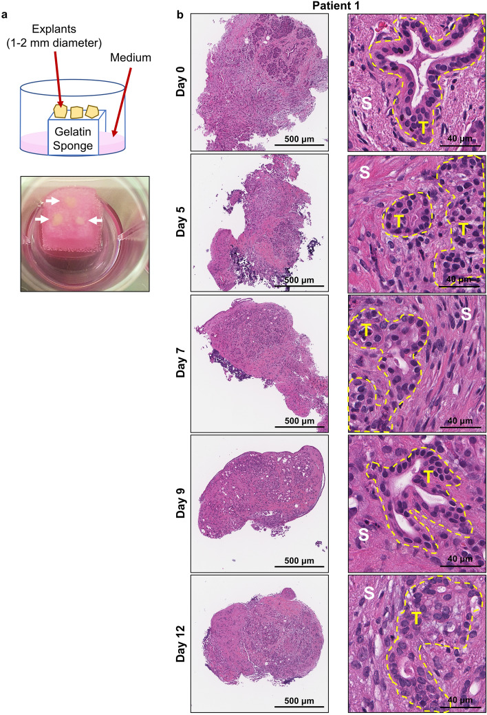
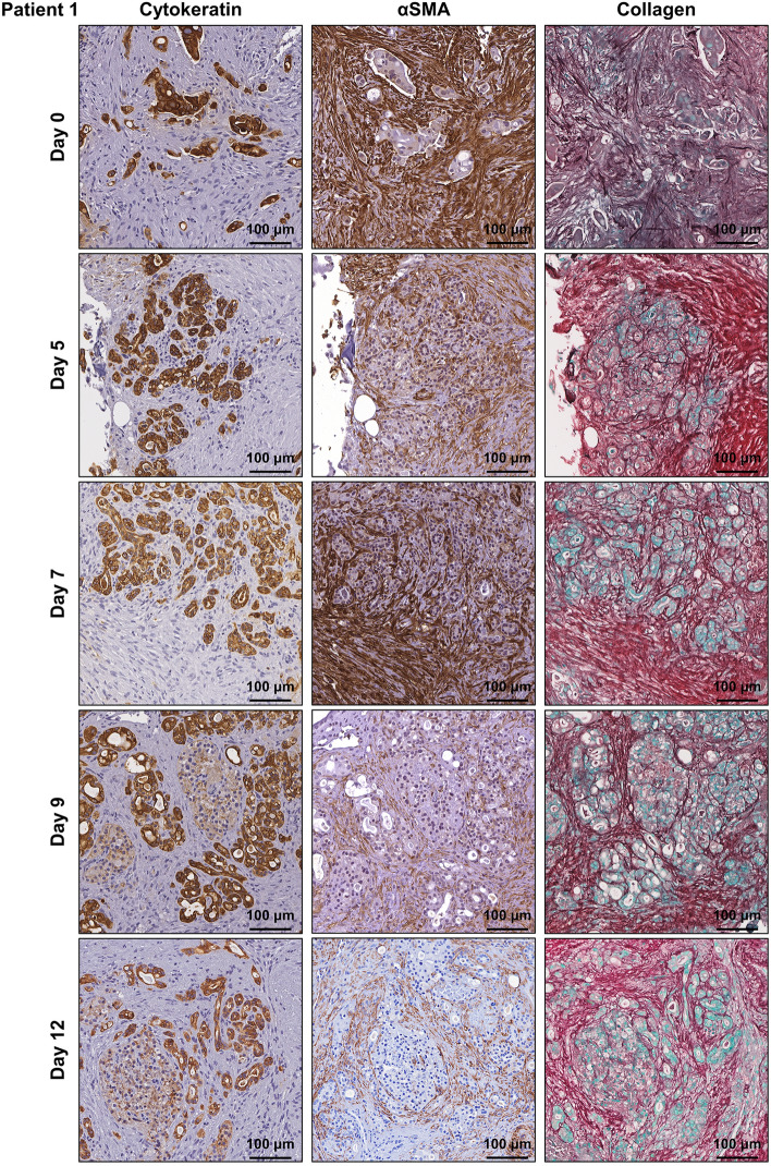
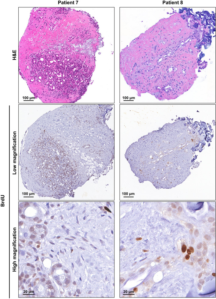
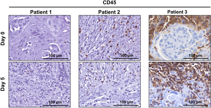
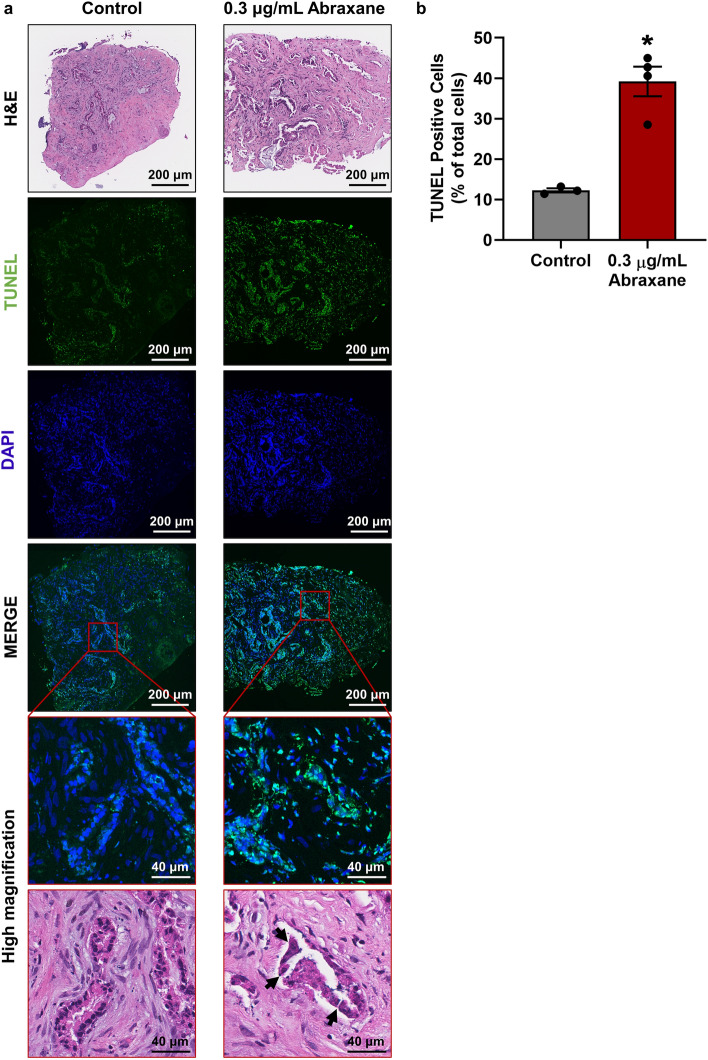
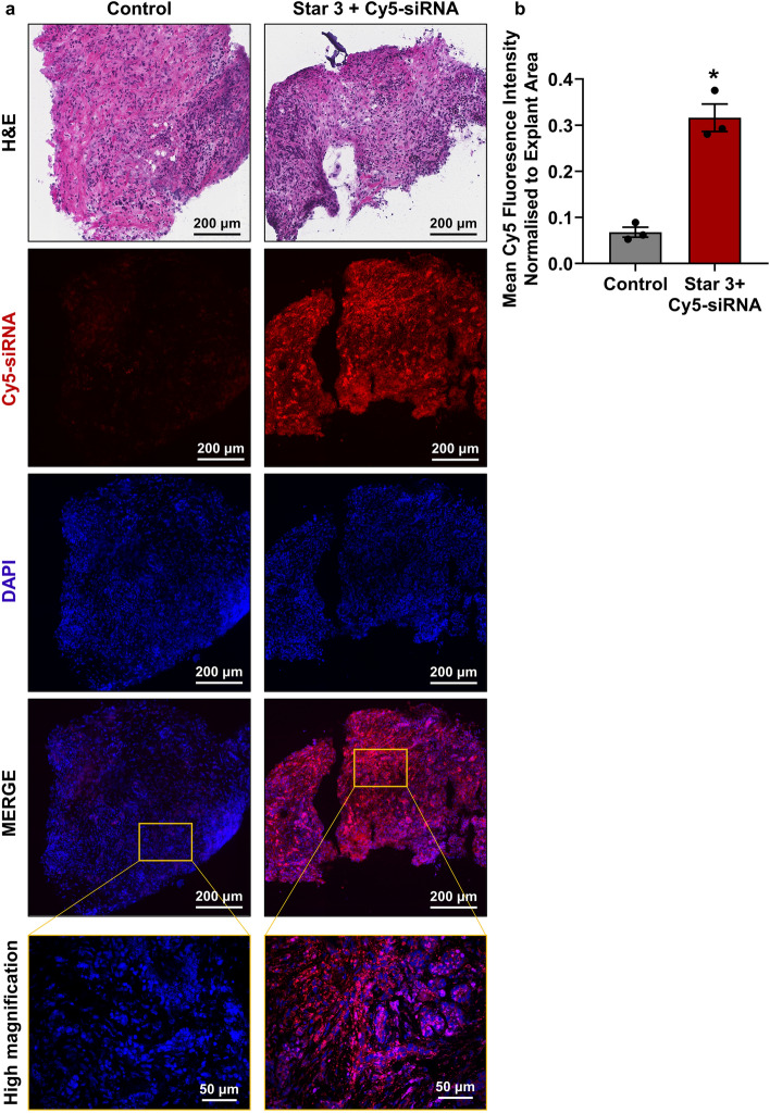
The poor prognosis of pancreatic ductal adenocarcinoma (PDAC) is attributed to the highly fibrotic stroma and complex multi-cellular microenvironment that is difficult to fully recapitulate in pre-clinical models. To fast-track translation of therapies and to inform personalised medicine, we aimed to develop a whole-tissue ex vivo explant model that maintains viability, 3D multicellular architecture, and microenvironmental cues of human pancreatic tumours. Patient-derived surgically-resected PDAC tissue was cut into 1-2 mm explants and cultured on gelatin sponges for 12 days. Immunohistochemistry revealed that human PDAC explants were viable for 12 days and maintained their original tumour, stromal and extracellular matrix architecture. As proof-of-principle, human PDAC explants were treated with Abraxane and we observed different levels of response between patients. PDAC explants were also transfected with polymeric nanoparticles + Cy5-siRNA and we observed abundant cytoplasmic distribution of Cy5-siRNA throughout the PDAC explants. Overall, our novel model retains the 3D architecture of human PDAC and has advantages over standard organoids: presence of functional multi-cellular stroma and fibrosis, and no tissue manipulation, digestion, or artificial propagation of organoids. This provides unprecedented opportunity to study PDAC biology including tumour-stromal interactions and rapidly assess therapeutic response to drive personalised treatment.


Conflict of interest statement

The authors declare no competing interests.

Figures

Figure 1
Figure 1
Tumour and stromal architecture are maintained in human patient derived pancreatic ductal adenocarcinoma explants for 12 days of culture. (a) Schematic showing the set-up of the ex vivo culture method and a representative photo of a gelatin sponge containing three PDAC explants in a well of a 24-well plate. White arrows point to the three explants on the gelatin sponge during culture. (b) Representative H&E images of patient 1 explants at low and high magnification from days 0–12. Tumour elements are outlined in yellow and compartments labelled as tumour (T) and stroma (S).
Figure 2
Figure 2
Characterisation of human patient derived pancreatic ductal adenocarcinoma explants cultured for 12 days. Immunohistochemistry was performed for cytokeratin, α-smooth muscle actin (αSMA), and collagen (picrosirius red/methyl green) on patient 1 explants from days 0–12.
Figure 3
Figure 3
Human patient derived pancreatic ductal adenocarcinoma tumour explants remain viable for 12 days of culture. Immunohistochemistry was performed for ki67, phospho-histone H3 (PHH3) and TUNEL on patient 1 explants from days 0–12. Insets show representative higher magnification views.
Figure 4
Figure 4
Patient derived pancreatic ductal adenocarcinoma tumour explants demonstrate positive bromodeoxyuridine (BrdU) staining after 12 days of culture. Patient 7 and 8 tumour explants were treated with 10 μM BrdU for 24 h prior to fixation at day 12. Immunohistochemistry demonstrated positive BrdU staining in both tumour and stromal cells in the day 12 explants.
Figure 5
Figure 5
Human patient derived pancreatic ductal adenocarcinoma tumour explants maintain p53 protein status after 12 days of culture. Immunohistochemistry was performed for p53 on day 0 and day 12 tumour explants from patients 1–6. Insets show representative higher magnification views.
Figure 6
Figure 6
CD45-positive lymphocytes remain viable for 5 days in human patient derived pancreatic ductal adenocarcinoma tumour explants. Immunohistochemistry was performed for lymphocyte marker CD45 on tumour explants from patients 1–3 at day 0 and day 5.
Figure 7
Figure 7
Testing of clinical therapeutics in human patient derived pancreatic ductal adenocarcinoma explants. (a) Tumour explants from patient 2 were treated with or without 0.3 μg/mL Abraxane on days 0, 3, 6 and 9, then fixed on day 12. TUNEL staining was performed to assess levels of cell death. Black arrows in H&E stained sections point to areas of ductal fragmentation in Abraxane treated explants. (b) Quantification of TUNEL positive cells using QuPath demonstrated increased cell death in Abraxane treated explants compared to untreated controls. A paired t-test was performed to compare TUNEL-positivity in control vs Abraxane treated explants from a single patient, with 3–4 explants per treatment from distinct regions of the patient’s tumour (represented by each dot in the bar graph). Bars represent mean ± S.E.M., *p = 0.0306.
Figure 8
Figure 8
Biodistribution of Star 3 nanoparticles in human patient derived pancreatic ductal adenocarcinoma explants. (a) Patient-derived explants from patient 13 were treated for 24 h with or without Star 3 polymeric nanoparticles coupled to Cy5-siRNA. Representative images show uptake of Cy5-siRNA throughout the entire explants. (b) Quantification of Cy5 mean fluorescence demonstrated significant uptake of Star 3 + Cy5-siRNA in treated explants compared to untreated controls. A paired t-test was performed to compare mean fluorescence normalised to explant area from a single patient, with 3 explants per treatment from distinct regions of the patient’s tumour (represented by each dot in the bar graph). Bars represent mean ± S.E.M., *p = 0.0176.

Similar articles

Cited by

References

    1. Siegel RL, Miller KD, Jemal A. Cancer statistics, 2020. CA Cancer J. Clin. 2020;70:7–30. doi: 10.3322/caac.21590. - DOI - PubMed
    1. Kamisawa T, Wood LD, Itoi T, Takaori K. Pancreatic cancer. Lancet. 2016;388:73–85. doi: 10.1016/s0140-6736(16)00141-0. - DOI - PubMed
    1. Wang Z, et al. Pancreatic cancer: Understanding and overcoming chemoresistance. Nat. Rev. Gastroenterol. Hepatol. 2011;8:27–33. doi: 10.1038/nrgastro.2010.188. - DOI - PubMed
    1. Erkan M, et al. The role of stroma in pancreatic cancer: Diagnostic and therapeutic implications. Nat. Rev. Gastroenterol. Hepatol. 2012;9:454–467. doi: 10.1038/nrgastro.2012.115. - DOI - PubMed
    1. Phillips, P. in Pancreatic Cancer and Tumour Microenvironment (ed Trivandrum) Chap. 3 (Transworld Research Network, 2012). - PubMed

Publication types

MeSH terms