

Early detection of diabetic retinopathy based on deep learning and ultra-wide-field fundus images
- PMID: 33479406
- PMCID: PMC7820327
- DOI: 10.1038/s41598-021-81539-3
Early detection of diabetic retinopathy based on deep learning and ultra-wide-field fundus images
Abstract
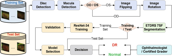
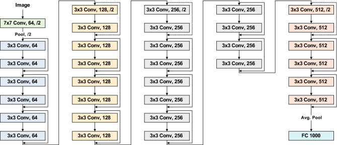
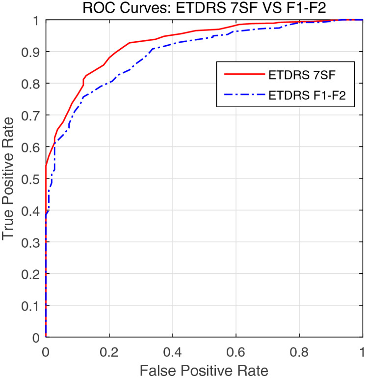
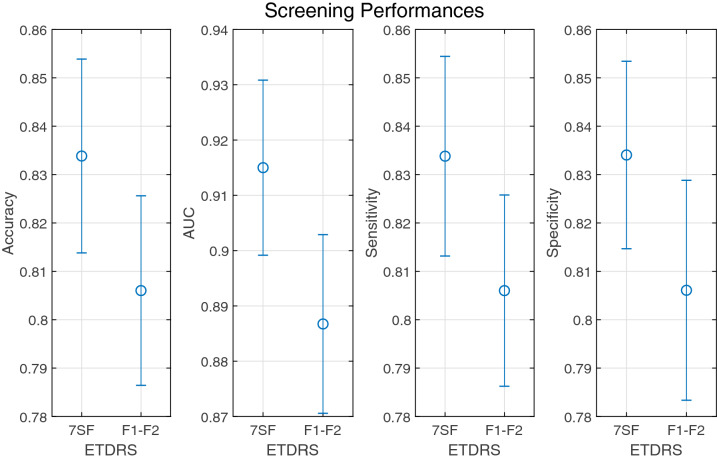
Visually impaired and blind people due to diabetic retinopathy were 2.6 million in 2015 and estimated to be 3.2 million in 2020 globally. Though the incidence of diabetic retinopathy is expected to decrease for high-income countries, detection and treatment of it in the early stages are crucial for low-income and middle-income countries. Due to the recent advancement of deep learning technologies, researchers showed that automated screening and grading of diabetic retinopathy are efficient in saving time and workforce. However, most automatic systems utilize conventional fundus photography, despite ultra-wide-field fundus photography provides up to 82% of the retinal surface. In this study, we present a diabetic retinopathy detection system based on ultra-wide-field fundus photography and deep learning. In experiments, we show that the use of early treatment diabetic retinopathy study 7-standard field image extracted from ultra-wide-field fundus photography outperforms that of the optic disc and macula centered image in a statistical sense.
Conflict of interest statement
The authors declare no competing interests.
Figures
References
-
- International Diabetes Federation. Diabetes atlas. IDF Diabetes Atlas, 9th edn. (International Diabetes Federation, Brussels, 2015).
-
- Thomas, R., Halim, S., Gurudas, S., Sivaprasad, S. & Owens, D. Idf diabetes atlas: A review of studies utilising retinal photography on the global prevalence of diabetes related retinopathy between 2015 and 2018. Diabetes Research and Clinical Practice, p. 107840 (2019). - PubMed
-
- Early Treatment Diabetic Retinopathy Study Research Group. Grading diabetic retinopathy from stereoscopic color fundus photographs-an extension of the modified airlie house classification: Etdrs report number 10. Ophthalmology98, 786–806 (1991). - PubMed
Publication types
MeSH terms
LinkOut - more resources
Full Text Sources
Other Literature Sources
Medical
