

Nanobody generation and structural characterization of Plasmodium falciparum 6-cysteine protein Pf12p
- PMID: 33480416
- PMCID: PMC7886318
- DOI: 10.1042/BCJ20200415
Nanobody generation and structural characterization of Plasmodium falciparum 6-cysteine protein Pf12p
Abstract
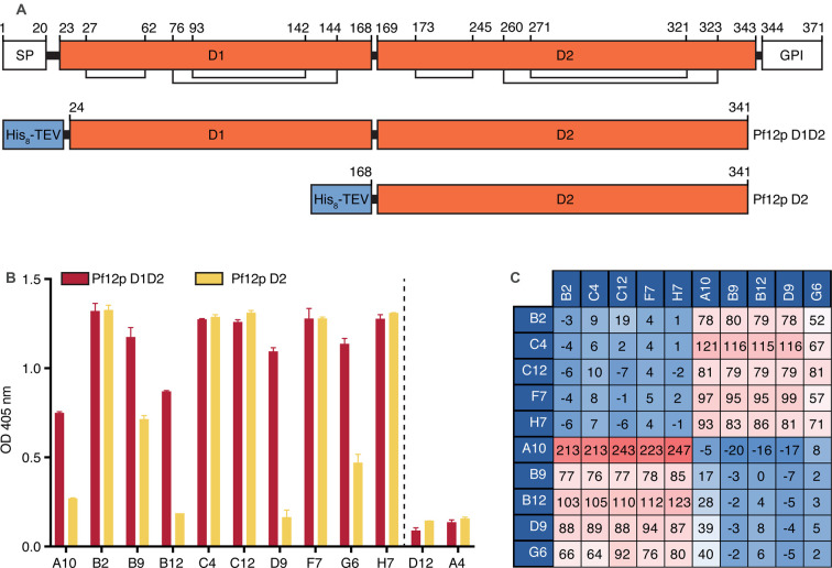
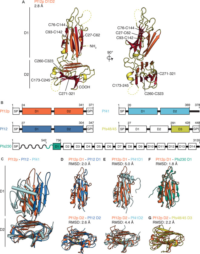
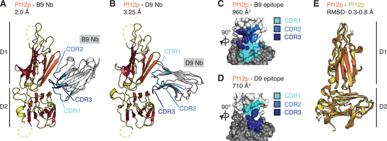
Surface-associated proteins play critical roles in the Plasmodium parasite life cycle and are major targets for vaccine development. The 6-cysteine (6-cys) protein family is expressed in a stage-specific manner throughout Plasmodium falciparum life cycle and characterized by the presence of 6-cys domains, which are β-sandwich domains with conserved sets of disulfide bonds. Although several 6-cys family members have been implicated to play a role in sexual stages, mosquito transmission, evasion of the host immune response and host cell invasion, the precise function of many family members is still unknown and structural information is only available for four 6-cys proteins. Here, we present to the best of our knowledge, the first crystal structure of the 6-cys protein Pf12p determined at 2.8 Å resolution. The monomeric molecule folds into two domains, D1 and D2, both of which adopt the canonical 6-cys domain fold. Although the structural fold is similar to that of Pf12, its paralog in P. falciparum, we show that Pf12p does not complex with Pf41, which is a known interaction partner of Pf12. We generated 10 distinct Pf12p-specific nanobodies which map into two separate epitope groups; one group which binds within the D2 domain, while several members of the second group bind at the interface of the D1 and D2 domain of Pf12p. Characterization of the structural features of the 6-cys family and their associated nanobodies provide a framework for generating new tools to study the diverse functions of the 6-cys protein family in the Plasmodium life cycle.
Keywords: 6-cysteine proteins; malaria; nanobody.
© 2021 The Author(s).
Conflict of interest statement
The authors declare that there are no competing interests associated with the manuscript.
Figures


Similar articles
-
Plasmodium 6-Cysteine Proteins: Functional Diversity, Transmission-Blocking Antibodies and Structural Scaffolds.Front Cell Infect Microbiol. 2022 Jul 8;12:945924. doi: 10.3389/fcimb.2022.945924. eCollection 2022. Front Cell Infect Microbiol. 2022. PMID: 35899047 Free PMC article. Review.
-
The Structure of Plasmodium falciparum Blood-Stage 6-Cys Protein Pf41 Reveals an Unexpected Intra-Domain Insertion Required for Pf12 Coordination.PLoS One. 2015 Sep 28;10(9):e0139407. doi: 10.1371/journal.pone.0139407. eCollection 2015. PLoS One. 2015. PMID: 26414347 Free PMC article.
-
Structure of the Pf12 and Pf41 heterodimeric complex of Plasmodium falciparum 6-cysteine proteins.FEMS Microbes. 2022 Feb 16;3:xtac005. doi: 10.1093/femsmc/xtac005. eCollection 2022. FEMS Microbes. 2022. PMID: 35308105 Free PMC article.
-
Structural and biochemical characterization of Plasmodium falciparum 12 (Pf12) reveals a unique interdomain organization and the potential for an antiparallel arrangement with Pf41.J Biol Chem. 2013 May 3;288(18):12805-17. doi: 10.1074/jbc.M113.455667. Epub 2013 Mar 19. J Biol Chem. 2013. PMID: 23511632 Free PMC article.
-
Disguising itself--insights into Plasmodium falciparum binding and immune evasion from the DBL crystal structure.Mol Biochem Parasitol. 2006 Jul;148(1):1-9. doi: 10.1016/j.molbiopara.2006.03.004. Epub 2006 Apr 4. Mol Biochem Parasitol. 2006. PMID: 16621067 Review.
Cited by
-
Plasmodium 6-Cysteine Proteins: Functional Diversity, Transmission-Blocking Antibodies and Structural Scaffolds.Front Cell Infect Microbiol. 2022 Jul 8;12:945924. doi: 10.3389/fcimb.2022.945924. eCollection 2022. Front Cell Infect Microbiol. 2022. PMID: 35899047 Free PMC article. Review.
-
Nanobodies against Cavin1 reveal structural flexibility and regulated interactions of its N-terminal coiled-coil domain.J Cell Sci. 2025 Apr 15;138(8):jcs263756. doi: 10.1242/jcs.263756. Epub 2025 Apr 28. J Cell Sci. 2025. PMID: 40260863 Free PMC article.
-
Rare catastrophes and evolutionary legacies: human germline gene variants in MLKL and the necroptosis signalling pathway.Biochem Soc Trans. 2022 Feb 28;50(1):529-539. doi: 10.1042/BST20210517. Biochem Soc Trans. 2022. PMID: 35166320 Free PMC article. Review.
-
Human coronavirus OC43 nanobody neutralizes virus and protects mice from infection.J Virol. 2024 Jun 13;98(6):e0053124. doi: 10.1128/jvi.00531-24. Epub 2024 May 6. J Virol. 2024. PMID: 38709106 Free PMC article.
-
Chemical Biology Tools To Investigate Malaria Parasites.Chembiochem. 2021 Jul 1;22(13):2219-2236. doi: 10.1002/cbic.202000882. Epub 2021 Mar 31. Chembiochem. 2021. PMID: 33570245 Free PMC article. Review.
References
-
- WHO (2019) World Malaria Report: 2019
-
- Arredondo, S.A., Swearingen, K.E., Martinson, T., Steel, R., Dankwa, D.A., Harupa, A.et al. (2018) The micronemal plasmodium proteins P36 and P52 Act in concert to establish the replication-permissive compartment within infected hepatocytes. Front. Cell Infect. Microbiol. 8, 413 10.3389/fcimb.2018.00413 - DOI - PMC - PubMed
Publication types
MeSH terms
Substances
Grants and funding
LinkOut - more resources
Full Text Sources
Other Literature Sources
Molecular Biology Databases