Review
. 2021 Apr;43(4):e2000286.
doi: 10.1002/bies.202000286. Epub 2021 Jan 22.

DNA topoisomerases: Advances in understanding of cellular roles and multi-protein complexes via structure-function analysis

Affiliations
Review

DNA topoisomerases: Advances in understanding of cellular roles and multi-protein complexes via structure-function analysis

Shannon J McKie et al. Bioessays. 2021 Apr.

Abstract

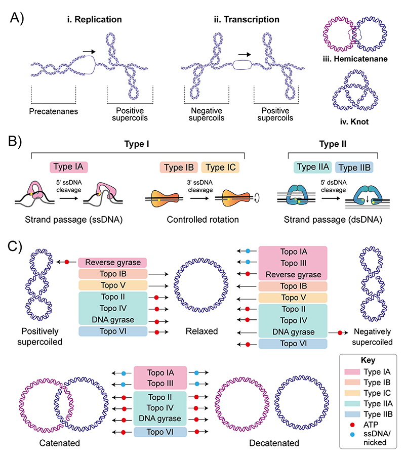
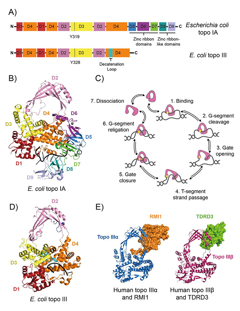
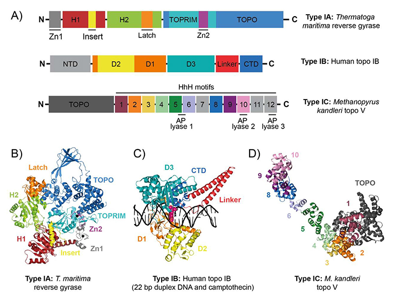
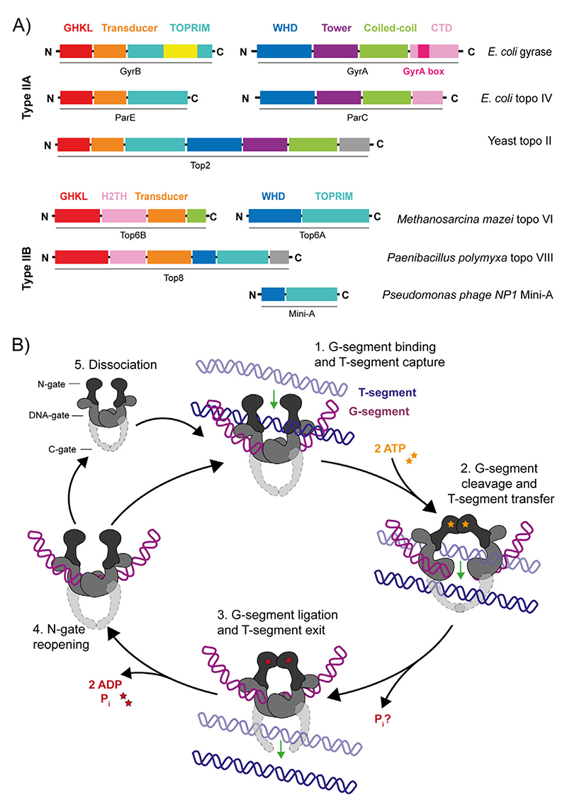
DNA topoisomerases, capable of manipulating DNA topology, are ubiquitous and indispensable for cellular survival due to the numerous roles they play during DNA metabolism. As we review here, current structural approaches have revealed unprecedented insights into the complex DNA-topoisomerase interaction and strand passage mechanism, helping to advance our understanding of their activities in vivo. This has been complemented by single-molecule techniques, which have facilitated the detailed dissection of the various topoisomerase reactions. Recent work has also revealed the importance of topoisomerase interactions with accessory proteins and other DNA-associated proteins, supporting the idea that they often function as part of multi-enzyme assemblies in vivo. In addition, novel topoisomerases have been identified and explored, such as topo VIII and Mini-A. These new findings are advancing our understanding of DNA-related processes and the vital functions topos fulfil, demonstrating their indispensability in virtually every aspect of DNA metabolism.

Keywords: DNA gyrase; DNA supercoiling; DNA topoisomerase; anti-cancer drugs; antibiotics.


Conflict of interest statement

Conflict of Interest

None declared.

Figures

Figure 1
Figure 1. DNA topology and DNA topoisomerase mechanisms.
(A) Topological consequences of DNA metabolism. i) During DNA replication, strand separation leads to positive supercoiling ahead of the advancing protein machinery, and precatenane formation behind. Precatenanes form as the newly-synthesised duplexes wrap around one and other, and, if not removed prior to complete of replication, catenated DNA molecules are formed. ii) During transcription, strand separation leads to positive supercoiling ahead of the advancing protein machinery, and negative supercoil formation behind. iii) Hemicatenanes are a possible end result of replication, in which the parental strands of the replicated duplexes remain base-paired. iv: DNA knotting can also occur as a result of DNA replication in which a DNA molecule is intramolecularly linked. (B) Summary of topo categories and mechanism. The topos are categorised based on whether they catalyse single- (type I) or double-stranded (type II) DNA breaks. The type I topos are further subdivided to type IA, IB and IC. Type IA form a transient covalent bond to the 5′ DNA phosphate and function via a strand passage mechanism. Type IB and IC form a transient covalent bond to the 3′ DNA phosphate and function via a controlled-rotation mechanism. Type II topos are further subdivided into type IIA and IIB. Both form a transient covalent bond to the 5′ DNA phosphate of both strands of the duplex and function via a strand-passage mechanism. (C) Summary of the topological manipulations performed by DNA topoisomerases, namely relaxation of positive and negative supercoils and decatenation. Type IA topos are colour-coded pink, type IB are orange, type IC are yellow, type IIA are green, and type IIB are blue. Requirement of ATP or ssDNA for activity is denoted using a red or blue circle, respectively
Figure 2
Figure 2. Type IA DNA topoisomerases.
(A) Protein domain organisation of Escherichia coli DNA topoisomerase IA (topo IA) and DNA topoisomerase III (topo III). Black vertical lines represent the active site tyrosines. (B) Crystal structure of E. coli topo I bound to ssDNA (PDB: 4RUL).[20] (C) Strand-passage mechanism for type IA topos. (1) topo binds G-segment ssDNA region, (2) the G-segment is cleaved. (3) The topo DNA-gate is opened, (4) which allows T-segment transfer through the cleaved G-strand. (5) The DNA gate is closed, (6) and the G-strand is re-ligated, changing the linking number by 1. (7) The topo can then go through another round of relaxation or dissociate from the DNA. Type IA topo (domains 1–4) is in pink, the active site tyrosine is yellow and the DNA is grey. (D) Crystal structure of E. coli topo III bound to ssDNA (PDB: 2O54).[26] (E) Crystal structures of human topo IIIα (blue) bound to RMI1(orange) (PDB: 4CGY),[39] and human topo IIIβ (magenta) bound to TDRD3 (green) (PDB: 5GVE).[60] For panels A, B and C, the topo I and III domains are colour coded as follows: D1 is red, D2 is pink, D3 is yellow, D4 is orange, D5 is marine blue, D6 is purple, D7 is green, D8 is teal, and D9 is light blue
Figure 3
Figure 3. Reverse gyrase (type IA), topo IB (type IB) and topo V (type IC).
(A) Protein domain organisation of Thermatoga maritima reverse gyrase, human DNA topoisomerase IB (topo IB), and Methanopyrus kandleri DNA topoisomerase V (topo V). (B) Crystal structure of T. maritima reverse gyrase (PDB: 4DDU).[71] (C) Crystal structure of human topo IB in a cleavage complex with a 22 bp duplex DNA and camptothecin (PDB: 1T8I).[220] (D) Crystal structure of M. kandleri topo V (PDB: 5HM5).[89]
Figure 4
Figure 4. Type II DNA topoisomerases: domain organisation and mechanism.
(A) Protein domain organisation for the type IIA topos: E. coli DNA gyrase, E. coli DNA topoisomerase IV (topo IV), yeast DNA topoisomerase II (topo II), Methanosarcina mazei DNA topoisomerase VI (topo VI), Paenibacillus polymyxa DNA topoisomerase VIII (plasmid-borne), and Pseudomonas phage NP1 Mini-A. (B) type II topo strand passage mechanism. (1) G-segment is bound at the DNA-gate and the T-segment is captured. (2) ATP binding stimulates dimerisation of the N-gate, the G-segment is cleaved and the T-segment is passed through the break. (3) The G-segment is re-ligated and T-segment exits through the C-gate. For type IIB topos, there is no C-gate so once the T-segment passes through the G-segment, it is released from the enzyme. (4) Dissociation of ADP and Pi allows N-gate opening, a scenario where the enzyme either remains bound to the G-segment, ready to capture a consecutive T-segment, or (5) dissociates from the G-segment.
Figure 5
Figure 5. Type IIA DNA topoisomerase structures.
(A) The E. coli gyrase GyrA CTD (PDB: 1ZI0)[129] and the E. coli topo IV ParC CTD (PDB: 1ZVT).[146] (B) CryoEM structure of full length E. coli gyrase complexed with a 130-bp DNA duplex and gepotidacin (PDB: 6RKW).[137] Colour coding for domains is as labelled in the figure with the second GyrA and GyrB coloured light grey and dark grey, respectively, and the DNA in black. (C) Crystal structure of Saccharomyces cerevisiae topo II with a 26 bp DNA duplex and ADPNP (PDB: 4GFH).[162] Colour coding of domains is as shown in the figure with second Top2 subunit coloured grey and the DNA in black
Figure 6
Figure 6. DNA topoisomerase VI (type IIB) structures.
(A) Crystal structure of Methanosarcina mazei topo VI (PDB: 2Q2E).[196] The domains are coloured as labelled in the figure on one TOP6A/Top6B heterodimer, with the second Top6A and Top6B coloured black and grey, respectively. (B) Crystal structure of Sulfolobus shibatae topo VI bound to radicicol (PDB: 2ZBK).[197] Colour coding is the same as in panel A except GHKL-bound radicicol is coloured yellow

References

    1. Watson JD, Crick FH. Genetical implications of the structure of deoxyribonucleic acid. Nature. 1953;171(4361):964–967. - PubMed
    1. Watson JD, Crick FHC. A structure for deoxyribose nucleic acid. Nature. 1953;171:737–738. - PubMed
    1. Postow L, Crisona NJ, Peter BJ, Hardy CD, Cozzarelli NR. Topological challenges to DNA replication: Conformations at the fork. Proc Natl Acad Sci U S A. 2001;98(15):8219–8226. doi: 10.1073/pnas.11100699898/15/8219. - DOI - PMC - PubMed
    1. Bates AD, Maxwell A. DNA Topology. Oxford University Press; Oxford: 2005.
    1. Racko D, Benedetti F, Goundaroulis D, Stasiak A. Chromatin loop extrusion and chromatin unknotting. Polymers (Basel) 2018;10(10) doi: 10.3390/polym10101126. - DOI - PMC - PubMed

Publication types

MeSH terms