. 2021 Apr 30;42(4):528-536.
doi: 10.1093/carcin/bgab005.

MicroRNA-374b inhibits breast cancer progression through regulating CCND1 and TGFA genes

Affiliations

MicroRNA-374b inhibits breast cancer progression through regulating CCND1 and TGFA genes

Yan Liu et al. Carcinogenesis. .

Abstract

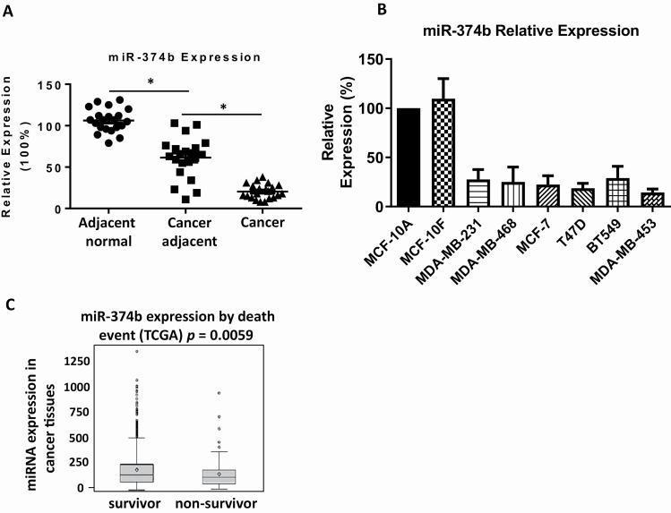
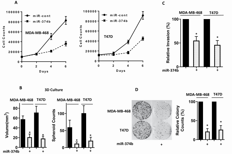
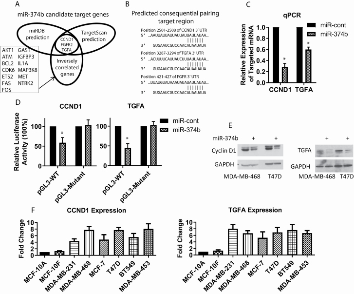
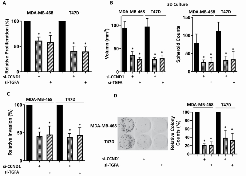
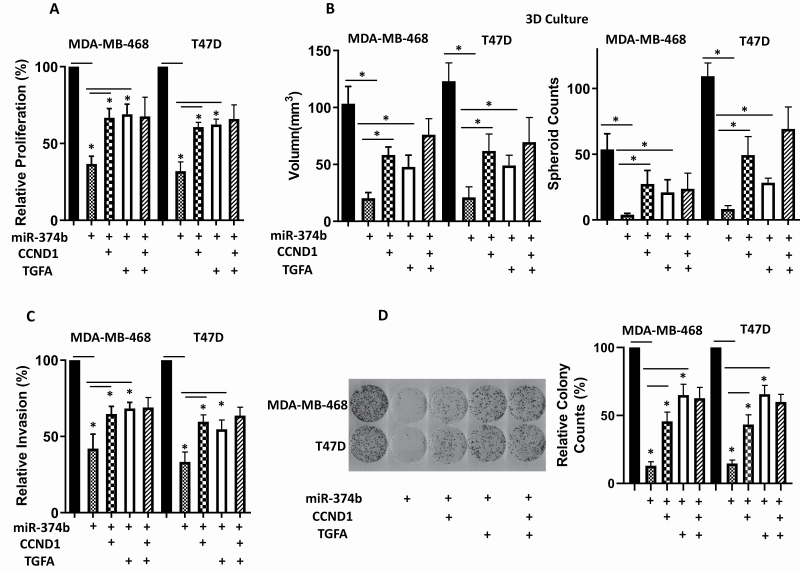
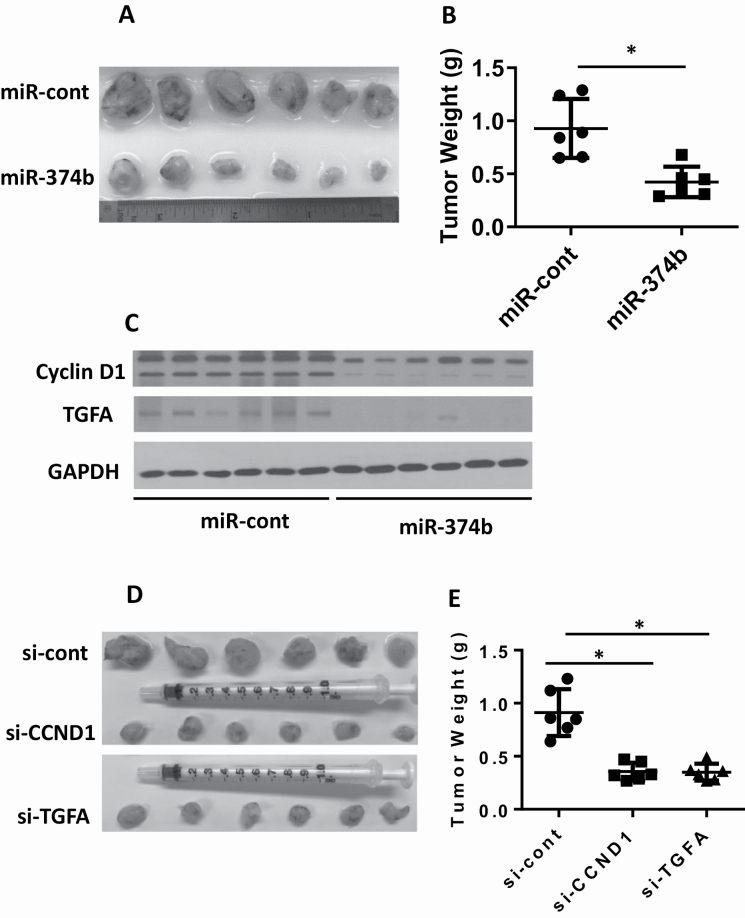
Emerging evidence indicates that microRNAs (miRNAs) play a critical role in breast cancer development. We recently reported that a higher expression of miR-374b in tumor tissues was associated with a better disease-free survival of triple-negative breast cancer (TNBC). However, the functional significance and molecular mechanisms underlying the role of miR-374b in breast cancer are largely unknown. In this current study, we evaluated the biological functions and potential mechanisms of miR-374b in both TNBC and non-TNBC. We found that miR-374b was significantly downregulated in breast cancer tissues, compared to adjacent tissues. MiR-374b levels were also lower in breast cancer cell lines, as compared to breast epithelial cells. In vitro and in vivo studies demonstrated that miR-374b modulates the malignant behavior of breast cancer cells, such as cell proliferation in 2D and 3D, cell invasion ability, colony-forming ability and tumor growth in mice. By using bioinformatics tools, we predicted that miR-374b plays a role in breast cancer cells through negatively regulating cyclin D1 (CCND1) and transforming growth factor alpha (TGFA). We further confirmed that CCND1 and TGFA contribute to the malignant behavior of breast cancer cells in vitro and in vivo. Our rescue experiments showed that overexpressing CCND1 or TGFA reverses the phenotypes caused by miR-374b overexpression. Taken together, our studies suggest that miR-374b modulates malignant behavior of breast cancer cells by negatively regulating CCND1 and TGFA genes. The newly identified miR-374b-mediated CCND1 and TGFA gene silencing may facilitate a better understanding of the molecular mechanisms of breast cancer progression.


Figures

Figure 1.
Figure 1.
MiR-374b is dysregulated in breast cancer tissues and breast cancer cell lines. (A) The levels of miR-374b in breast cancer tissues, paired adjacent cancer tissues and paired adjacent normal tissues were measured by quantitative real-time PCR. (B) The levels of miR-374b in human breast epithelial cells (MCF-10A and MCF-10F) and breast cancer cells (MDA-MB-231, MDA-MB-468, MCF-7, T47D, BT549 and MDA-MB-453) were examined by real-time PCR. (C) MiR-374b expression stratified by death event in TCGA (1058 patients). *P < 0.05.
Figure 2.
Figure 2.
Overexpressing miR-374b disrupts the malignant behavior of breast cancer cells. MiR-374b was transfected into MDA-MB-468 and T47D cells. Forty-eight hours post-transfection, functional assays were performed. (A) Cell proliferation was measured by MTT assay at different time points. (B) Multicellular tumor spheroids formation using 3D cell culture was examined. (C) Cell migration ability was measured by trans-well migration assay. (D) Colony-forming ability was examined by clonogenic assay. Three independent experiments were performed, and the data were presented as mean ± SD. *P < 0.05.
Figure 3.
Figure 3.
MiR-374b negatively regulates the expression of CCND1 and TGFA in breast cancer cells. (A) CCND1 and TGFA were predicted as direct targets of miR-374b (TargetScan and miRDB). (B) The potential miR-374b targeting sites at the 3′ UTR of CCND1, TGFA and FGFR2 genes. (C) The expression of CCND1 and TGFA mRNA in miR-374b-transfected MDA-MB-468 cells was measured by real-time PCR. (D) Relative luciferase level was examined after luciferase reporter plasmids with CCND1 or TGFA 3′ UTR (WT or mutant), or control reporter plasmid miR-374b or miR-control, were co-transfected in MDA-MB-468 cells. (E) The levels of CCND1 and TGFA in miR-374b-transfected cells were examined by western blotting. (F) The levels of CCND1 and TGFA mRNA in human breast epithelial cells and cancer cells were analyzed by real-time PCR. *P < 0.05.
Figure 4.
Figure 4.
Knocking down CCND1 or TGFA disrupts the malignant behavior of breast cancer cells. MDA-MB-468 and T47D cells were transfected with si-CCND1, si-TGFA or siRNA negative control. Forty-eight hours post-transfection, functional assays were performed. (A) Cell proliferation was measured by MTT assay at 96 h post-transfection. (B) Multicellular tumor spheroids formation using 3D cell culture was examined. (C) Cell migration ability was analyzed by trans-well migration assay. (D) Colony-forming ability was investigated by clonogenic assay. Three independent experiments were performed, and the data were presented as mean ± SD. *P < 0.05.
Figure 5.
Figure 5.
Overexpressing CCND1 or TGFA partially disrupts the effects of overexpressing miR-374b on the malignant behavior of breast cancer cells. MiR-374b was co-transfected into breast cancer cells with expression construct of CCND1 or TGFA, or both. (A) Cell proliferation was measured by MTT assay at 96 h post-transfection. (B) Multicellular tumor spheroids formation using 3D cell culture was examined. (C) Cell migration ability was analyzed by trans-well migration assay. (D) Colony-forming ability was investigated by clonogenic assay. Three independent experiments were performed, and the data were presented as mean ± SD. *P < 0.05.
Figure 6.
Figure 6.
Overexpressing miR-374b represses tumor growth in vivo through regulating CCND1 and TGFA. MDA-MB-468 stable cells were generated to constitutively express miR-374b or miR-control. (A and B) MDA-MB-468 stable cells expressing miR-374b or miR-control were injected subcutaneously into nude mice. After 5 weeks, tumor weight was evaluated. (C) Total protein lysates were extracted from tumor xenografts and the levels of cyclin D1 and TGFA were evaluated by western blotting. MDA-MB-468 stable cells were generated to constitutively express si-CCND1, si-TGFA or si-control. (D and E) 5 × 10⁶ cells were injected subcutaneously into nude mice. After 5 weeks, tumor weight was evaluated.

Similar articles

Cited by

References

    1. Brennecke, J., et al. (2003) bantam encodes a developmentally regulated microRNA that controls cell proliferation and regulates the proapoptotic gene hid in Drosophila. Cell, 113, 25–36. - PubMed
    1. Chen, C.Z., et al. (2004) MicroRNAs modulate hematopoietic lineage differentiation. Science, 303, 83–86. - PubMed
    1. Schratt, G.M., et al. (2006) A brain-specific microRNA regulates dendritic spine development. Nature, 439, 283–289. - PubMed
    1. Bartel, D.P. (2009) MicroRNAs: target recognition and regulatory functions. Cell, 136, 215–233. - PMC - PubMed
    1. Croce, C.M. (2009) Causes and consequences of microRNA dysregulation in cancer. Nat. Rev. Genet., 10, 704–714. - PMC - PubMed