. 2022 Jul;42(5):1373-1384.
doi: 10.1007/s10571-020-01022-x. Epub 2021 Jan 22.

Insulin-Like Growth Factor-1 Enhances Motoneuron Survival and Inhibits Neuroinflammation After Spinal Cord Transection in Zebrafish

Affiliations

Insulin-Like Growth Factor-1 Enhances Motoneuron Survival and Inhibits Neuroinflammation After Spinal Cord Transection in Zebrafish

Liping Zhao et al. Cell Mol Neurobiol. 2022 Jul.

Abstract

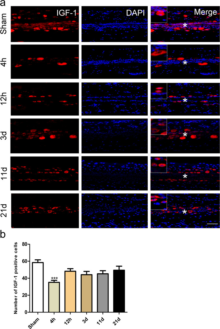
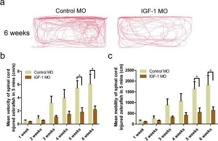
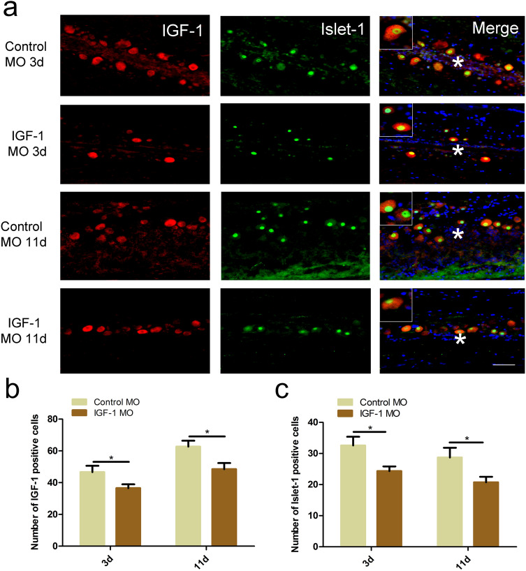
Insulin-like growth factor-1 (IGF-1) is a neurotrophic factor produced locally in the central nervous system which can promote axonal regeneration, protect motoneurons, and inhibit neuroinflammation. In this study, we used the zebrafish spinal transection model to investigate whether IGF-1 plays an important role in the recovery of motor function. Unlike mammals, zebrafish can regenerate axons and restore mobility in remarkably short period after spinal cord transection. Quantitative real-time PCR and immunofluorescence showed decreased IGF-1 expression in the lesion site. Double immunostaining for IGF-1 and Islet-1 (motoneuron marker)/GFAP (astrocyte marker)/Iba-1 (microglia marker) showed that IGF-1 was mainly expressed in motoneurons and was surrounded by astrocyte and microglia. Following administration of IGF-1 morpholino at the lesion site of spinal-transected zebrafish, swimming test showed retarded recovery of mobility, the number of motoneurons was reduced, and increased immunofluorescence density of microglia was caused. Our data suggested that IGF-1 enhances motoneuron survival and inhibits neuroinflammation after spinal cord transection in zebrafish, which suggested that IGF-1 might be involved in the motor recovery.

Keywords: Insulin-like growth factor-1; Motoneuron; Neuroinflammation; Spinal cord transection; Zebrafish.


Conflict of interest statement

The authors declare that they have no conflict of interest.

Figures

Fig. 1
Fig. 1
Time course of insulin-like growth factor-1 (IGF-1) mRNA expression after spinal transection. Quantitative real-time PCR showed the expression of IGF-1 mRNA in the spinal cord tissue within 1.5 mm upstream and downstream of the injury site after spinal transection. Decreased expression was observed at 3 days, 11 days, and 21 days after spinal cord transection compared to the sham injury group (*P < 0.05, **P < 0.01, independent samples t test; n = 8). Values represent mean ± SEM
Fig. 2
Fig. 2
Time course of insulin-like growth factor-1 (IGF-1) protein expression after spinal transection. a IGF-1 protein expression in spinal cord within 1.5 mm upstream and downstream of the injury site were examined by immunofluorescence at 5 time points after spinal transection. Cytoplasmic expression of IGF-1 protein was confirmed. Decreased IGF-1-positive cells were observed along the central canal at 4 h after spinal transection. *Indicates the central canal, scale bar = 50 μm. b Number of IGF-1-positive cells was quantified with ImageJ software. The lesion-induced decrease of IGF-1 expression achieved significance at 4 h post injury (*P < 0.05, **P < 0.01, ***P < 0.001, Kruskal–Wallis test; n = 3). Values represent mean ± SEM
Fig. 3
Fig. 3
Cytoplasmic expression of insulin-like growth factor-1 (IGF-1) in motoneurons. Double-immunostaining staining of IGF-1 with Islet-1 in longitudinal sections (1.5 mm upstream and downstream from lesion site) showed that IGF-1 was located in the cytoplasmic of motoneurons at 3 days post injury (n = 3). *Indicates the central canal. Scale bar = 50 μm
Fig. 4
Fig. 4
IGF-1-positive motoneurons were surrounded by GFAP-positive astrocytes. Double-immunostaining staining of IGF-1 with GFAP in longitudinal sections (1.5 mm upstream and downstream from lesion site) showed that IGF-1 was surrounded by GFAP-positive astrocytes at 3 days post lesion (n = 3). *Indicates the central canal. Scale bar = 50 μm
Fig. 5
Fig. 5
Increased fluorescence density of microglia after spinal transection. a Double immunostaining of insulin-like growth factor-1 (IGF-1) with Iba-1 in longitudinal sections (1.5 mm upstream and downstream from lesion site) showed that IGF-1 was surrounded by microglia. *Indicates the central canal, scale bar = 50 μm. b Immunofluorescence density of Iba-1 was quantified with ImageJ software. The lesion-induced increase of immunofluorescence density of microglia achieved significance at 12 h, 3 days, 11 days, and 21 days after spinal transection (*P < 0.05, **P < 0.01, ***P < 0.001, one-way ANOVA with an LSD post hoc assay; n = 3). Values represent mean ± SEM
Fig. 6
Fig. 6
The insulin-like growth factor-1 (IGF-1) morpholino (MO) significantly reduced IGF-1 mRNA expression. a Schematic illustration showed the spinal cord transection site and morpholino treatment site. b cDNA was amplified from RNA isolated from IGF-1 knockdown morphants, control MO-injected embryos, and non-injected embryos. RT-PCR was conducted using the primers in exons 3 and 4 for IGF-1. c and d Enrichment of un-spliced fragments was showed on the representative agarose gel. Reduction of un-spliced fragment on IGF-1 MO injected embryos was observed at both 24 h post-fertilization (b) and 48 h post-fertilization (c)
Fig. 7
Fig. 7
Knockdown of insulin-like growth factor-1 (IGF-1) expression inhibited motor recovery. a Locus diagram of the control morpholino (MO) and IGF-1 MO groups at 6 weeks. b and c Mean velocity and total distance moved in 5 min of zebrafish treated with IGF-1 MO and control MO after spinal cord transection. At 5 and 6 weeks post injury, mean velocity and total distance moved were significantly reduced in IGF-1 MO group compared with control MO group (*P < 0.05, Mann–Whitney U test was used for mean velocity and mean mobility between two groups at 5 weeks, independent samples t test was used for mean velocity and mean mobility between two groups at 6 weeks; n = 9). Values represent mean ± SEM
Fig. 8
Fig. 8
Islet-1-positive motoneurons decrease after insulin-like growth factor-1 (IGF-1) morpholino (MO) treated. a Double immunostaining of IGF-1 with Islet-1 was performed in IGF-1 MO group and control MO group. *Indicates the central canal, scale bar = 50 μm. b and c The number of IGF-1-positive cells and motoneurons was both decreased in IGF-1 MO group compare with control MO group (*P < 0.05, Mann–Whitney U test; n = 3). Values represent mean ± SEM
Fig. 9
Fig. 9
Insulin-like growth factor-1 (IGF-1) morpholino (MO) treatment induced increased immunofluorescence density of microglia. a Double immunostaining of IGF-1 and Iba-1 showed decreased IGF-1 expression in IGF-1 MO group compared with control MO group at 3 days and 11 days after injury. *Indicates the central canal, scale bar = 50 μm. b Increased fluorescence density of Iba-1 in IGF-1 MO group at 3 days and 11 days after injury was observed compared with control MO group (*P < 0.05, **P < 0.01, independent samples t test; n = 3). Values represent mean ± SEM

Similar articles

Cited by

References

    1. Aghanoori M-R, Smith DR, Shariati-Ievari S, Ajisebutu A, Nguyen A, Desmond F, Jesus CHA, Zhou X, Calcutt NA, Aliani M, Fernyhough P (2019) Insulin-like growth factor-1 activates AMPK to augment mitochondrial function and correct neuronal metabolism in sensory neurons in type 1 diabetes. Mol Metab 20:149–165. 10.1016/j.molmet.2018.11.008 - PMC - PubMed
    1. Becker T, Bernhardt RR, Reinhard E, Wullimann MF, Tongiorgi E, Schachner M (1998) Readiness of zebrafish brain neurons to regenerate a spinal axon correlates with differential expression of specific cell recognition molecules. J Neurosci 18(15):5789–5803 - PMC - PubMed
    1. Bluthe RM, Kelley KW, Dantzer R (2006) Effects of insulin-like growth factor-I on cytokine-induced sickness behavior in mice. Brain Behav Immun 20(1):57–63. 10.1016/j.bbi.2005.02.003 - PMC - PubMed
    1. Bollaerts I, Van Houcke J, Andries L, De Groef L, Moons L (2017) Neuroinflammation as fuel for axonal regeneration in the injured vertebrate central nervous system. Mediat Inflamm 2017:9478542. 10.1155/2017/9478542 - PMC - PubMed
    1. Boucher SE, Hitchcock PF (1998) Insulin-related growth factors stimulate proliferation of retinal progenitors in the goldfish. J Comp Neurol 394(3):386–394 - PubMed

Substances

LinkOut - more resources