

Sphenoidal artery: review of the literature and analysis of a dissected arterially injected fetal orbit
- PMID: 33481129
- PMCID: PMC7897611
- DOI: 10.1007/s00276-020-02663-9
Sphenoidal artery: review of the literature and analysis of a dissected arterially injected fetal orbit
Abstract
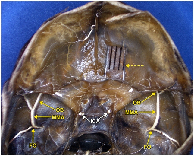
Purpose: The sphenoidal artery is considered a component of the complex and dangerous arterial anastomoses of the human orbitocranial region, particularly with the advent of interventional neuroimaging. The objective of this publication was to analyze the various descriptions of the sphenoidal artery in the literature as related to relevant photographs of a dissected arterially injected fetal middle cranial fossa and orbit.
Methods: Publications dealing with middle meningeal-ophthalmic arterial anastomoses, focusing on the sphenoidal artery, were reviewed. A relevant dissection of a fetal specimen was analyzed.
Results: The literature dealing with the sphenoidal artery is at times not in agreement. The nomenclature and anatomy of its passage through the superior orbital fissure or Hyrtl canal have variable descriptions. Photographs of the skull base of a dissected arterially injected fetal specimen show bilateral prominent orbital branches of the middle meningeal arteries. These branches entered both orbits in a course similar to the diagrammatic representations of the sphenoidal artery, and give rise to several major intraorbital arteries. This study provides the only photographic image in the literature of this variation in a human fetal anatomic dissection.
Conclusions: Review of the literature dealing with the sphenoidal artery shows inconsistent nomenclature and conflicting descriptions of its anastomotic connections, and varying evolutionary and embryologic theories. Analysis of the dissected fetal skull base indicates that the sphenoidal artery is not a distinct artery but just a middle meningeal orbital arterial branch, an important component of the complex and dangerous arterial anastomoses of the human orbitocranial region.
Keywords: Hyrtl canal; Middle meningeal artery; Ophthalmic artery, orbit; Sphenoidal artery; Superior orbital fissure.
Conflict of interest statement
The authors declared no potential conflicts of interest with respect to the research, authorship and/or publication of this article.
Figures

Similar articles
-
A microsurgical study of the anatomy and course of the ophthalmic artery and its possibly dangerous anastomoses.J Neurosurg. 2007 Jan;106(1):142-50. doi: 10.3171/jns.2007.106.1.142. J Neurosurg. 2007. PMID: 17236500
-
The cranio-orbital foramen, the groove on the lateral wall of the human orbit, and the orbital branch of the middle meningeal artery.Clin Anat. 2005 Jan;18(1):10-4. doi: 10.1002/ca.20020. Clin Anat. 2005. PMID: 15597374
-
Absence of foramen spinosum and abnormal middle meningeal artery in cranial series.Anthropol Anz. 2012 Jul;69(3):351-66. doi: 10.1127/0003-5548/2012/0165. Anthropol Anz. 2012. PMID: 22928356
-
The revised anatomy of the canals connecting the orbit with the cranial cavity.Orbit. 2017 Apr;36(2):110-117. doi: 10.1080/01676830.2017.1279662. Epub 2017 Mar 3. Orbit. 2017. PMID: 28388344 Review.
-
Orbital vascular anatomy.Eye (Lond). 2006 Oct;20(10):1130-44. doi: 10.1038/sj.eye.6702377. Eye (Lond). 2006. PMID: 17019411 Review.
References
-
- Clay C, Vignaud J. Vascularisation de l’orbite. Encyclopedie Medico-Chirugicale (Paris) 1971;10:1–12.
Publication types
MeSH terms
Grants and funding
LinkOut - more resources
Full Text Sources
Other Literature Sources
