

Novel typing of iliac vein compression in asymptomatic individuals evaluated by contrast enhanced CT
- PMID: 33481132
- PMCID: PMC8273055
- DOI: 10.1007/s00276-021-02678-w
Novel typing of iliac vein compression in asymptomatic individuals evaluated by contrast enhanced CT
Abstract
Purpose: Compression of the iliac vein between the iliac artery and lumbosacral vertebra can cause iliac vein compression syndrome (IVCS). The purpose of this study is to assess compression characteristics and establish a new sub-typing in asymptomatic IVCS individuals using contrast-enhanced CT.
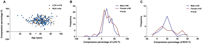
Methods: A retrospective analysis of abdomen contrast-enhanced CT images from 195 asymptomatic subjects with iliac vein compressed was investigated. Patients had no history of venous pathology, and images were collected from June 2018 to January 2019. Qualitative and quantitative characteristics of compression were examined including the location, pattern, minor diameter, area, and the percentage compression on an orthogonal section by the post-processing of multiple planar reconstruction and volume rendering.
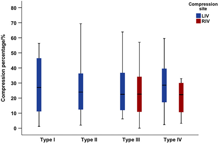
Results: There were 107 females and 88 males with age range 18-92 years. The most common site of iliac vein compression was localized to the left common iliac vein (LCIV) (178/195, 91.3%). Notably, four compression types (type I-IV) were established according to the compression location, with type II being the most common. The four compression types had differences in the upper limit and fluctuation range of compression. It was found that the average level of iliac vein compression was below 25%. The compression degree of the left common iliac vein in type II was relatively concentrated, and the upper limit of compression was close to 70%.
Conclusion: Asymptomatic iliac vein compression was categorized according to compression location. The proposal of four types might help clinicians to predict which IVCS patients would benefit from interventional therapy.
Keywords: Asymptomatic diseases; Compression type; Iliac vein; Iliac vein compression syndrome; May-Thurner syndrome; Tomography; X-ray computed.
© 2021. The Author(s).
Conflict of interest statement
The authors declare that they have no conflict of interests.
Figures


Similar articles
-
Dual compression is not an uncommon type of iliac vein compression syndrome.Int J Cardiovasc Imaging. 2017 Sep;33(9):1277-1285. doi: 10.1007/s10554-017-1112-4. Epub 2017 Mar 13. Int J Cardiovasc Imaging. 2017. PMID: 28289992
-
May-Thurner syndrome: can it be diagnosed by a single MR venography study?Diagn Interv Radiol. 2013 Jan-Feb;19(1):44-8. doi: 10.4261/1305-3825.DIR.5939-12.1. Epub 2012 Jul 16. Diagn Interv Radiol. 2013. PMID: 22801870
-
Compression of the left common iliac vein in asymptomatic subjects and patients with left iliofemoral deep vein thrombosis.J Vasc Interv Radiol. 2008 Mar;19(3):366-70; quiz 371. doi: 10.1016/j.jvir.2007.09.007. J Vasc Interv Radiol. 2008. PMID: 18295695
-
Diagnosis and management of iliac vein compression syndrome.J Vasc Nurs. 2005 Mar;23(1):10-7; quiz 18-9. doi: 10.1016/j.jvn.2004.12.001. J Vasc Nurs. 2005. PMID: 15741959 Review.
-
A Comprehensive Review of the Pathophysiology and Clinical Importance of Iliac Vein Obstruction.Eur J Vasc Endovasc Surg. 2020 Jul;60(1):118-125. doi: 10.1016/j.ejvs.2020.03.020. Epub 2020 Apr 17. Eur J Vasc Endovasc Surg. 2020. PMID: 32312667 Review.
Cited by
-
Impact of anatomical features of non-thrombotic left iliac venous compression on the development of venous leg ulcers based on CT venography.Abdom Radiol (NY). 2025 Aug;50(8):3876-3884. doi: 10.1007/s00261-024-04772-0. Epub 2025 Jan 27. Abdom Radiol (NY). 2025. PMID: 39869216
-
Iliac vein compression syndrome by lumbar degenerative changes is associated with deep vein thrombosis after total knee arthroplasty.Arch Orthop Trauma Surg. 2023 Sep;143(9):5833-5842. doi: 10.1007/s00402-023-04811-3. Epub 2023 Feb 17. Arch Orthop Trauma Surg. 2023. PMID: 36799994
-
CFD Study of the Effect of the Angle Pattern on Iliac Vein Compression Syndrome.Bioengineering (Basel). 2023 Jun 5;10(6):688. doi: 10.3390/bioengineering10060688. Bioengineering (Basel). 2023. PMID: 37370619 Free PMC article.
-
Frequency of left common iliac vein compression in asymptomatic adolescents and young adults.J Vasc Surg Venous Lymphat Disord. 2025 Jun 20;13(6):102282. doi: 10.1016/j.jvsv.2025.102282. Online ahead of print. J Vasc Surg Venous Lymphat Disord. 2025. PMID: 40545194 Free PMC article.
References
MeSH terms
Substances
Grants and funding
LinkOut - more resources
Full Text Sources
Other Literature Sources
