. 2021:2251:143-156.
doi: 10.1007/978-1-0716-1142-5_10.

Characterization of Protein-Phospholipid/Membrane Interactions Using a "Membrane-on-a-Chip" Microfluidic System

Affiliations

Characterization of Protein-Phospholipid/Membrane Interactions Using a "Membrane-on-a-Chip" Microfluidic System

Calvin Yeager et al. Methods Mol Biol. 2021.

Abstract

It is now clear that organelles of a mammalian cell can be distinguished by phospholipid profiles, both as ratios of common phospholipids and by the absence or presence of certain phospholipids. Organelle-specific phospholipids can be used to provide a specific shape and fluidity to the membrane and/or used to recruit and/or traffic proteins to the appropriate subcellular location and to restrict protein function to this location. Studying the interactions of proteins with specific phospholipids using soluble derivatives in isolation does not always provide useful information because the context in which the headgroups are presented almost always matters. Our laboratory has shown this circumstance to be the case for a viral protein binding to phosphoinositides in solution and in membranes. The system we have developed to study protein-phospholipid interactions in the context of a membrane benefits from the creation of tailored membranes in a channel of a microfluidic device, with a fluorescent lipid in the membrane serving as an indirect reporter of protein binding. This system is amenable to the study of myriad interactions occurring at a membrane surface as long as a net change in surface charge occurs in response to the binding event of interest.

Keywords: Fluorescence; Label-free; Microfluidics; Phosphatidylinositol 4-phosphate; Phosphoinositides; Pleckstrin Homology domain; Supported lipid bilayer; pH modulation.


Figures

Fig. 1
Fig. 1
The membrane-on-a-chip device is a microfluidic platform. (a) The device is composed of three major components: a PDMS block with an imprinted micropattern; a borosilicate glass coverslip; and a microscope slide. Holes are punched in the PDMS block for inlet and outlet tubing. (b) The micropattern provides eight microchannels for establishment of supported lipid bilayers (SLBs). Inlet and outlet holes should be punched at the black circles. (c) The microfluidic device loaded onto an epifluorescence microscope. Outlet tubing feeds into a petri dish; inlet tubing brings in running buffer from gravity flow. The microchannels are capable of being fluorescently excited from an inverted microscope setup
Fig. 2
Fig. 2
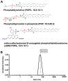
Small unilamellar vesicles (SUVs) can be formed from lipids of interest. (a) Our experiments contain phosphatidylcholine (POPC), phosphatidylinositol-4-phosphate (PI4P), and ortho-sulforhodamine B conjugated to phosphatidylethanolamine (oSRB-POPE). Depending on the user’s need, SUVs from other lipid mixtures can be used. (b) Measurement of hydrodynamic radius of 92 mol% POPC, 7.5 mol% PI4P, and 0.5 mol% oSRB-POPE vesicles. Dynamic light scattering (DLS) can be used to assure SUVs within their associated hydrodynamic radius size range of 15–90 nm
Fig. 3
Fig. 3
The oSRB-POPE probe is fluorescent and pH sensitive. (a) Primary data of the microfluidic channels. All channels in the “Before” image are at pH 7.0. A range of different pH buffers are introduced to those channels to highlight the dynamic range of 0.5 mol% oSRB-POPE in the “After” image. (b) Fluorescence quantitation of panel a with a line tool. (c) Plotted absolute fluorescence versus pH values. The pKa of the oSRB-POPE probe in 92 mol% POPC, 7.5 mol% PI4P, and 0.5 mol% oSRB-POPE is 6.5
Fig. 4
Fig. 4
The membrane-on-a-chip can detect protein—membrane interactions. (a) Protein is added to the microchannels, and it interacts with lipids in the bilayer. The protein carries counterions, which will interact with oSRB-POPE and impact the fluorescence values of the channel. (b) Increasing concentrations of a PV 3C derivative bind to saturation on a 92 mol% POPC, 7.5 mol% PI4P, and 0.5 mol% oSRB-POPE membrane. (c) Plotted values from panel b and Langmuir isotherm fit. The Kd,app was determined to be 3.0 ± 0.2 μM

References

    1. Lemmon MA (2008) Membrane recognition by phospholipid-binding domains. Nat Rev Mol Cell Biol 9(2):99–111. 10.1038/nrm2328 - DOI - PubMed
    1. Kutateladze TG (2010) Translation of the phosphoinositide code by PI effectors. Nat Chem Biol 6(7):507–513. 10.1038/nchembio.390 - DOI - PMC - PubMed
    1. Jung H, Robison AD, Cremer PS (2009) Detecting protein-ligand binding on supported bilayers by local pH modulation. J Am Chem Soc 131(3):1006–1014. 10.1021/ja804542p - DOI - PMC - PubMed
    1. Narayan K, Lemmon MA (2006) Determining selectivity of phosphoinositide-binding domains. Methods 39(2):122–133. 10.1016/j.ymeth.2006.05.006 - DOI - PMC - PubMed
    1. Scott JL, Musselman CA, Adu-Gyamfi E, Kutateladze TG, Stahelin RV (2012) Emerging methodologies to investigate lipid-protein interactions. Integr Biol (Camb) 4(3):247–258. 10.1039/c2ib00143h - DOI - PMC - PubMed

LinkOut - more resources