. 2021 Feb 3;69(4):1272-1282.
doi: 10.1021/acs.jafc.0c06827. Epub 2021 Jan 22.

First Characterization of the Formation of Anthocyanin-Ge and Anthocyanin-B Complexes through UV-Vis Spectroscopy and Density Functional Theory Quantum Chemical Calculations

Affiliations

First Characterization of the Formation of Anthocyanin-Ge and Anthocyanin-B Complexes through UV-Vis Spectroscopy and Density Functional Theory Quantum Chemical Calculations

Laura Estévez et al. J Agric Food Chem. .

Abstract

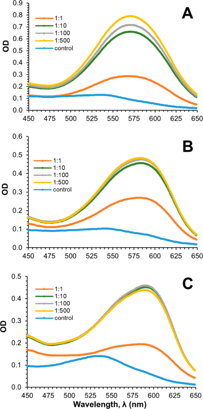
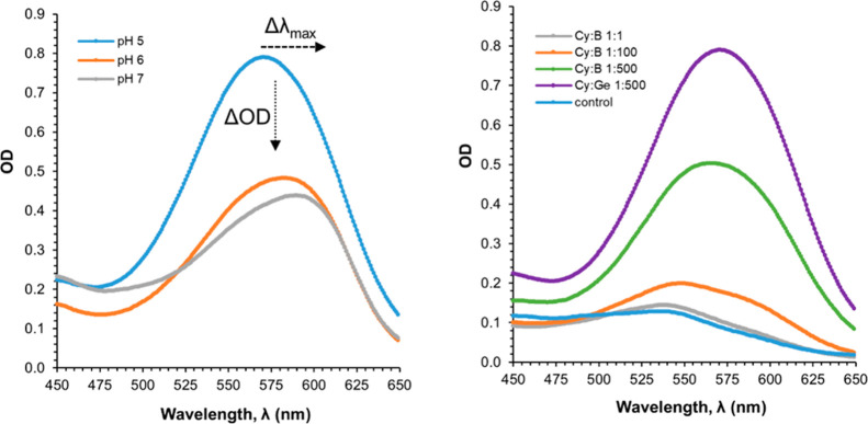
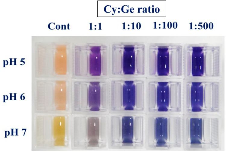
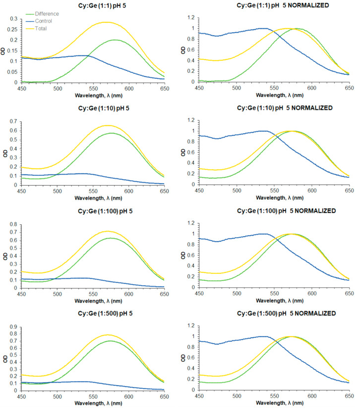
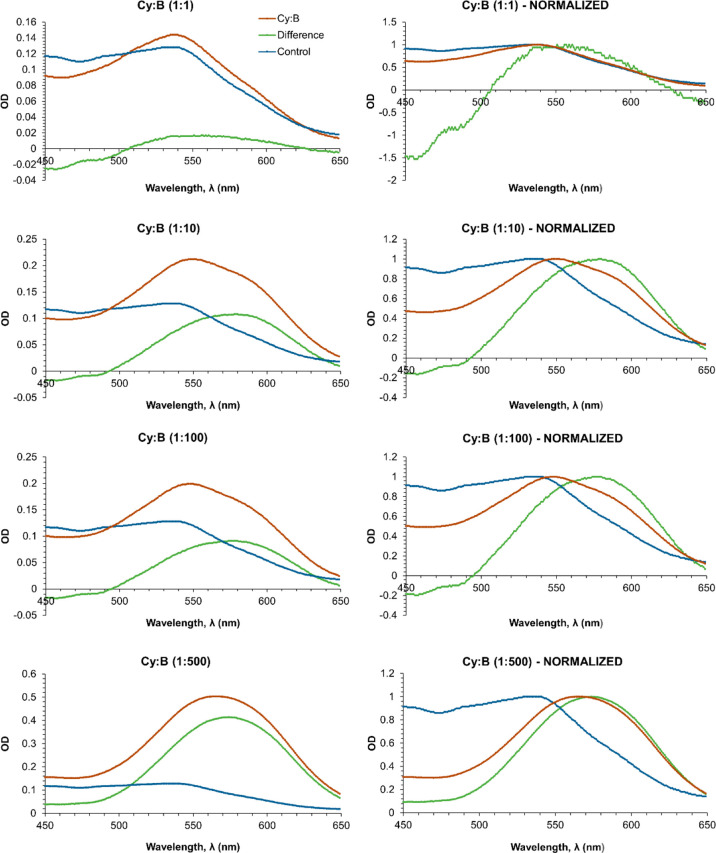
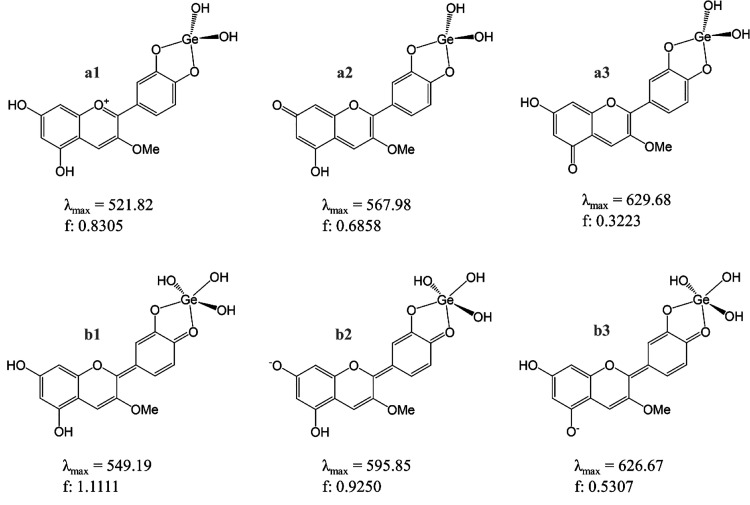
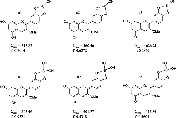
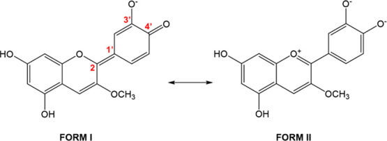
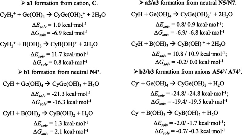
The occurrence of anthocyanin (ACN) and metal (Me) complexes has been widely supported by many research works while the possibility that ACNs bind to metalloids (Mds) is yet to be proven. Here, metalloids (H3BO3 for B; GeO2 for Ge) were added to cyanidin-based solutions at pH 5, 6, and 7 and ACN-Md stoichiometric ratios of 1:1, 1:10, 1:100, and 1:500, and UV-vis transmittance spectroscopy as well as density functional theory (DFT) calculations were performed to test this hypothesis. Ge and B addition caused bathochromic and hyperchromic shifts on ACN UV-vis spectra, particularly pronounced at pH 5 and a 1:500 (ACN:Md) ratio. ACN-Me complexation reactions have been evaluated where Ge showed a higher capability to bind to ACNs than B. Among the complexes envisioned, those labeled as b1, b2, and b3 feature UV-vis spectra compatible with experiments. The combination of experimental and computational data offers for the first time evidence of the formation of ACN-Md complexes.

Keywords: anthocyanin−metalloid complex; bathochromic shift; hyperchromic effect; molecular absorption simulations; molecular modeling.


Conflict of interest statement

The authors declare no competing financial interest.

Figures

Scheme 1
Scheme 1. pH Equilibria for the Cyanidin-3-OMe Model System
Estimated (see text) pKa values (pKa1(cation→N4′) 4.8 and pKa2(N4′→A54′) 7.2) and energy difference between tautomers of the anion form, ΔEA54′A74′ (in bold, kcal·mol–1), are shown.
Figure 1
Figure 1
UV–vis spectra for the various cyanidin-based anthocyanins complexed with Ge at different ratios (Cy:Ge). Cy:Ge ratios investigated: 1:1, 1:10, 1:100, and 1:500 at (A) pH 5, (B) pH 6, and (C) pH 7.
Figure 2
Figure 2
UV–vis spectra for (left) cyanidin-based anthocyanins complexed with Ge at a 1:500 ratio, at the pH values explored (5, 6, and 7) and (right) with B at various Cy:B ratios and comparison with Cy:Ge (1:500) at pH 5.
Figure 3
Figure 3
Change in visual appearance of the reaction mixtures of cyanidin-based anthocyanins isolated from Red Rubin sweet basil leaves and Ge ions added with different stoichiometric ratios (1:1, 1:10, 1:100, 1:500; Cy:Ge) at pH 5, 6, and 7.
Figure 4
Figure 4
UV–vis spectra of cyanidin-based anthocyanins solutions containing Ge at the various Cy:Ge ratios explored (yellow curves), at pH 5. The curves (green) resulting from subtracting the control solution (blue curves) spectrum (left) are also shown. Normalized curves (right) are also shown to more clearly note bathochromic shifts.
Figure 5
Figure 5
UV–vis spectra of cyanidin-based anthocyanins solutions containing B at the various Cy:B ratios explored (red curves), at pH 5. The curves (green) resulting from subtracting the control solution (blue curves) spectrum (left) are also shown. Normalized curves (right) are also shown to more clearly note bathochromic shifts.
Scheme 2
Scheme 2. Cyanidin-Based Complexes with Ge (Cy–Ge) Explored with Color Forms of Cyanin
a1 is obtained from the Cy cation, C; a2, a3, and b1 are from N7, N5, and N4′, respectively; and b2 and b3 are from A74′ and A54′, respectively. The corresponding wavelength of maximum absorbance, λmax, nm, and oscillator strength, f, are shown for each complex.
Scheme 3
Scheme 3. Cyanidin-Based Complexes with B (Cy–B) Explored with Color Forms of Cyanin
a1 is obtained from the Cy cation, C; a2, a3, and b1 are from N7, N5, and N4′, respectively, and b2 and b3 are from A74′ and A54′, respectively. The corresponding wavelength of maximum absorbance, λmax, nm, and oscillator strength, f, are shown for each complex.
Scheme 4
Scheme 4. Main Resonance Forms of the Aglycon within the Metalloid Complex
Scheme 5
Scheme 5. Main Complexation Reactions Involving Color Forms of Cyanin
CyH2+ stands for the cation form, CyH stands for any of the three neutral tautomers, and Cy stands for any of the anions, giving rise to a1a3 and b1b3 complexes. The ΔEsolv and ΔGsolv (kcal·mol–1) values for corresponding processes are shown.

References

    1. Grotewold E. The genetics and biochemistry of floral pigments. Annu. Rev. Plant Biol. 2006, 57, 761–780. 10.1146/annurev.arplant.57.032905.105248. - DOI - PubMed
    1. Landi M.; Tattini M.; Gould K. S. Multiple functional roles of anthocyanins in plant-environment interactions. Environ. Exp. Bot. 2015, 119, 4–17. 10.1016/j.envexpbot.2015.05.012. - DOI
    1. Yoshida K.; Mori M.; Kondo T. Blue flower color development by anthocyanins: from chemical structure to cell physiology. Nat. Prod. Rep. 2009, 26, 884.10.1039/b800165k. - DOI - PubMed
    1. Hatier J. H. B.; Gould K. S.. Anthocyanin function in vegetative organs. In Anthocyanins: Biosynthesis, Functions, and Applications; Gould K. S., Davies K. M., Winefield C., Eds.; Springer: New York, 2009; pp 1–20.
    1. Ellestad G. A. Structure and chiroptical properties of supramolecular flower pigments. Chirality 2006, 18, 134–144. 10.1002/chir.20228. - DOI - PubMed