Comparative Study
. 2021 Jan 22;16(1):e0245795.
doi: 10.1371/journal.pone.0245795. eCollection 2021.

A comparative study on the use of microscopy in pharmacology and cell biology research

Affiliations
Comparative Study

A comparative study on the use of microscopy in pharmacology and cell biology research

Agatha M Reigoto et al. PLoS One. .

Abstract

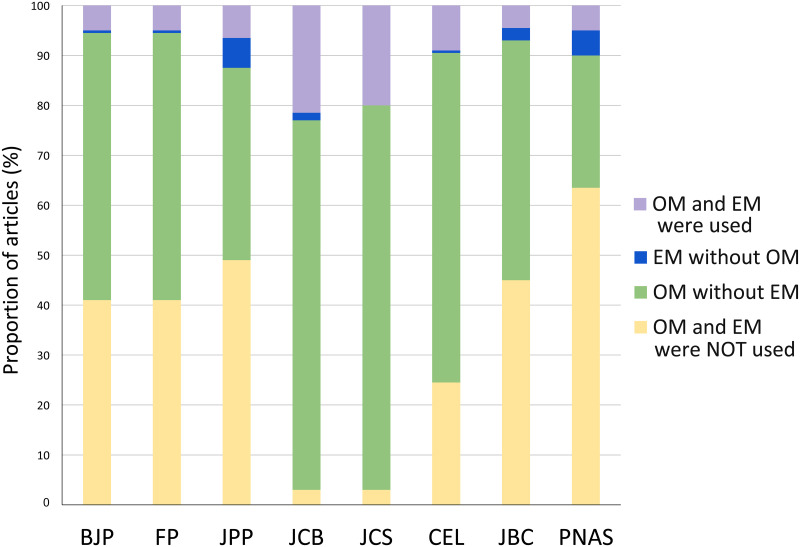
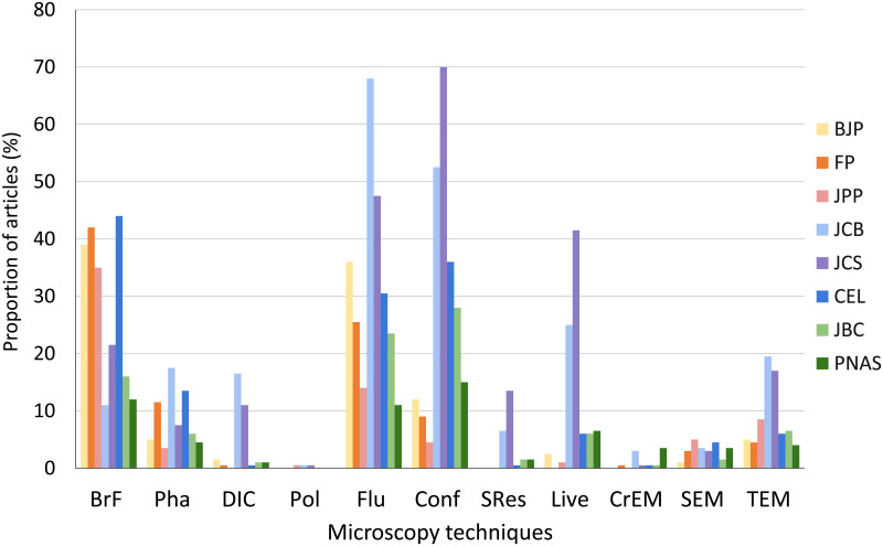
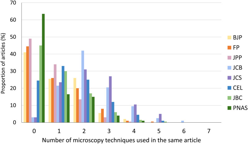
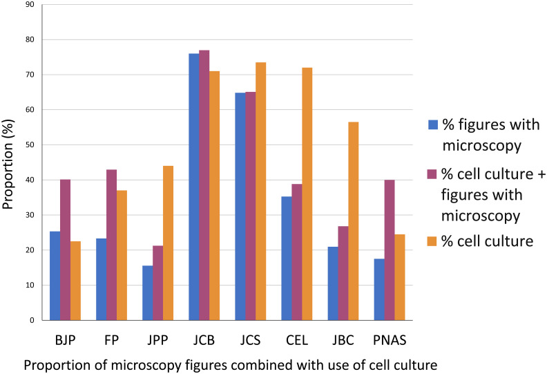
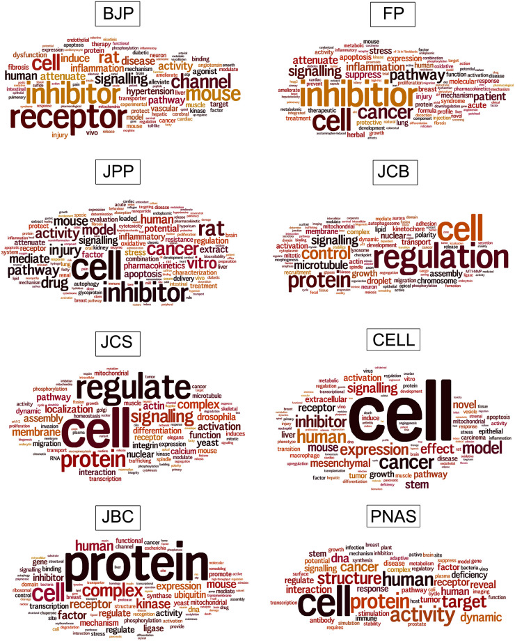
Microscopy is the main technique to visualize and study the structure and function of cells. The impact of optical and electron microscopy techniques is enormous in all fields of biomedical research. It is possible that different research areas rely on microscopy in diverse ways. Here, we analyzed comparatively the use of microscopy in pharmacology and cell biology, among other biomedical sciences fields. We collected data from articles published in several major journals in these fields. We analyzed the frequency of use of different optical and electron microscopy techniques: bright field, phase contrast, differential interference contrast, polarization, conventional fluorescence, confocal, live cell imaging, super resolution, transmission and scanning electron microscopy, and cryoelectron microscopy. Our analysis showed that the use of microscopy has a distinctive pattern in each research area, and that nearly half of the articles from pharmacology journals did not use any microscopy method, compared to the use of microscopy in almost all the articles from cell biology journals. The most frequent microscopy methods in all the journals in all areas were bright field and fluorescence (conventional and confocal). Again, the pattern of use was different: while the most used microscopy methods in pharmacology were bright field and conventional fluorescence, in cell biology the most used methods were conventional and confocal fluorescence, and live cell imaging. We observed that the combination of different microscopy techniques was more frequent in cell biology, with up to 6 methods in the same article. To correlate the use of microscopy with the research theme of each article, we analyzed the proportion of microscopy figures with the use of cell culture. We analyzed comparatively the vocabulary of each biomedical sciences field, by the identification of the most frequent words in the articles. The collection of data described here shows a vast difference in the use of microscopy among different fields of biomedical sciences. The data presented here could be valuable in other scientific and educational contexts.


Conflict of interest statement

The authors have declared that no competing interests exist.

Figures

Fig 1
Fig 1. Comparative analysis of the proportion of articles that uses microscopy techniques in biomedical sciences journals.
Analysis of the presence of optical (OM) and electron (EM) microscopy methodologies in all articles published in 2019 in the journals: British Journal of Pharmacology (BJP), Journal of Pharmacy and Pharmacology (JPP), Frontiers in Pharmacology (FP), Journal of Cell Biology (JCB), Journal of Cell Science (JCS), Cells (CEL), Journal of Biological Chemistry (JBC) and Proceedings of the National Academy of Sciences (PNAS). N = 200 articles analyzed in each scientific journal.
Fig 2
Fig 2. Comparative analysis of the proportion of use of different optical and electron microscopy techniques in biomedical sciences journals.
We analyzed in each article the use of 8 types of optical microscopy techniques: bright field (BrF), phase contrast (Pha), differential interference contrast (DIC), polarization (Pol), conventional fluorescence (Flu), confocal fluorescence (Conf), super resolution (SRes), and live cell imaging (Live); and 3 types of electron microscopy techniques: transmission (TEM), scanning (SEM) and cryo-EM (CrEM). Data were collected from articles published in 2019 in the biomedical sciences journals: British Journal of Pharmacology (BJP), Journal of Pharmacy and Pharmacology (JPP), Frontiers in Pharmacology (FP), Journal of Cell Biology (JCB), Journal of Cell Science (JCS), Cells (CEL), Journal of Biological Chemistry (JBC) and Proceedings of the National Academy of Sciences (PNAS). N = 200 articles analyzed in each scientific journal.
Fig 3
Fig 3. Comparative analysis of the number of microscopy techniques used in articles from biomedical sciences journals.
Data were collected from articles published in 2019 in the biomedical sciences journals: British Journal of Pharmacology (BJP), Journal of Pharmacy and Pharmacology (JPP), Frontiers in Pharmacology (FP), Journal of Cell Biology (JCB), Journal of Cell Science (JCS), Cells (CEL), Journal of Biological Chemistry (JBC) and Proceedings of the National Academy of Sciences (PNAS). N = 200 articles analyzed in each scientific journal.
Fig 4
Fig 4. Comparative analysis of the percentage of articles figures with microscopy and cell culture in biomedical sciences journals.
The percentage of figures containing microscopy images is shown in blue bars, the percentage of articles containing cell culture is shown in orange bars and the percentage of articles containing cell culture and microscopy is shown in magenta. Data were collected from articles published in 2019 in the biomedical sciences journals: British Journal of Pharmacology (BJP), Journal of Pharmacy and Pharmacology (JPP), Frontiers in Pharmacology (FP), Journal of Cell Biology (JCB), Journal of Cell Science (JCS), Cells (CEL), Journal of Biological Chemistry (JBC) and Proceedings of the National Academy of Sciences (PNAS). N = 200 articles analyzed in each scientific journal.
Fig 5
Fig 5. Comparative analysis of the vocabulary of biomedical sciences journals.
Word clouds were generated using the titles of articles. The clouds give greater prominence to words that appear more frequently in the source text. Data were collected from articles published in 2019 in the biomedical sciences journals: British Journal of Pharmacology (BJP), Journal of Pharmacy and Pharmacology (JPP), Frontiers in Pharmacology (FP), Journal of Cell Biology (JCB), Journal of Cell Science (JCS), Cells (CELL), Journal of Biological Chemistry (JBC) and Proceedings of the National Academy of Sciences (PNAS). N = 200 articles analyzed in each scientific journal.

Similar articles

Cited by

References

    1. Morris JD, Payne CK. Microscopy and cell biology: new methods and new questions. Annu Rev Phys Chem. 2019; 70: 199–218. 10.1146/annurev-physchem-042018-052527 - DOI - PubMed
    1. Albuquerque PC, Fonseca BPF, Girard-Dias W, Zicker F, de Souza W, Miranda K. Mapping the Brazilian microscopy landscape: A bibliometric and network analysis. Micron 2019; 116: 84–92. 10.1016/j.micron.2018.10.005 - DOI - PubMed
    1. Hooke R. Micrographia: or, some physiological descriptions of minute bodies made by magnifying glasses. 1st ed London: J. Martyn and J. Allestry; 1665.
    1. Usaj MM, Styles EB, Verster AJ, Friesen H, Boone C, Andrews BJ. High-Content Screening for Quantitative Cell Biology. Trends Cell Biol. 2016; 26: 598–611. 10.1016/j.tcb.2016.03.008 - DOI - PubMed
    1. Inoué S. Polarization microscopy. Curr Protoc Cell Biol. 2002; Chapter 4: Unit 4.9. 10.1002/0471143030.cb0409s13 - DOI - PubMed

Publication types

LinkOut - more resources