

Incidence and characterization of acute pulmonary embolism in patients with SARS-CoV-2 pneumonia: A multicenter Italian experience
- PMID: 33481902
- PMCID: PMC7822531
- DOI: 10.1371/journal.pone.0245565
Incidence and characterization of acute pulmonary embolism in patients with SARS-CoV-2 pneumonia: A multicenter Italian experience
Abstract
Background and aims: Several studies reported a high incidence of pulmonary embolism (PE) among patients with severe acute respiratory syndrome coronavirus-2 (SARS-CoV-2) infection, but detailed data about clinical characteristics, risk factors of these patients and prognostic role of PE are still lacking. We aim to evaluate the occurrence of pulmonary embolism among patients with SARS-CoV-2 infection, and to describe their risk factors, clinical characteristics, and in-hospital clinical outcomes.
Methods: This is a multicenter Italian study including 333 consecutive SARS-CoV-2 patients admitted to seven hospitals from February 22 to May 15, 2020. All the patients underwent computed tomography pulmonary angiography (CTPA) for PE detection. In particular, CTPA was performed in case of inadequate response to high-flow oxygen therapy (Fi02≥0.4 to maintain Sp02≥92%), elevated D-dimer (>0.5μg/mL), or echocardiographic signs of right ventricular dysfunction. Clinical, laboratory and radiological data were also analyzed.
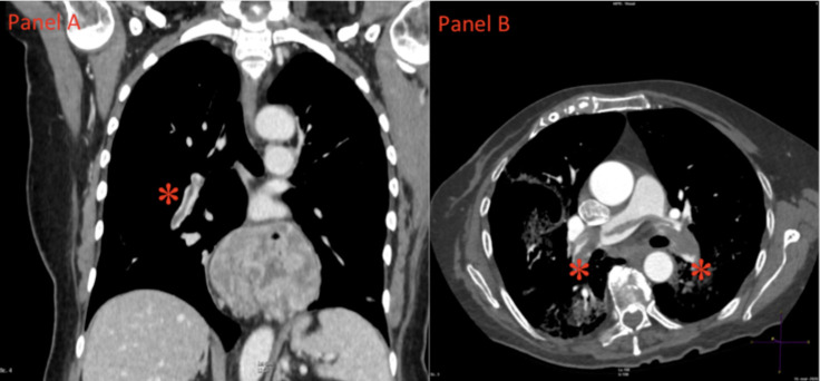
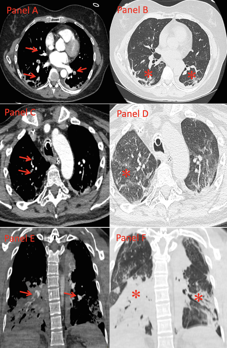
Results: Among 333 patients with laboratory confirmed SARS-CoV-2 pneumonia and undergoing CTPA, PE was detected in 109 (33%) cases. At CTPA, subsegmental, segmental, lobar and central thrombi were detected in 31 (29%), 50 (46%), 20 (18%) and 8 (7%) cases, respectively. In-hospital death occurred in 29 (27%) patients in the PE-group and in 47 (21%) patients in the non-PE group (p = 0.25). Patients in PE-group had a low rate of traditional risk factors and deep vein thrombosis was detected in 29% of patients undergoing compression ultrasonography. In 71% of cases with documented PE, the thrombotic lesions were located in the correspondence of parenchymal consolidation areas.
Conclusions: Despite a low rate of risk factors for venous thromboembolism, PE is present in about 1 out 3 patients with SARS-CoV-2 pneumonia undergoing CTPA for inadequate response to oxygen therapy, elevated D-dimer level, or echocardiographic signs of right ventricular dysfunction. In most of the cases, the thromboses were located distally in the pulmonary tree and were mainly confined within pneumonia areas.
Conflict of interest statement
The authors also have declared that no competing interests exist.
Figures
References
Publication types
MeSH terms
Substances
LinkOut - more resources
Full Text Sources
Other Literature Sources
Medical
Miscellaneous
