. 2021 Jan-Jun:296:100309.
doi: 10.1016/j.jbc.2021.100309. Epub 2021 Jan 19.

Poly(ADP-ribose) polymerase 1 regulates mitochondrial DNA repair in an NAD-dependent manner

Affiliations

Poly(ADP-ribose) polymerase 1 regulates mitochondrial DNA repair in an NAD-dependent manner

Geoffrey K Herrmann et al. J Biol Chem. 2021 Jan-Jun.

Abstract

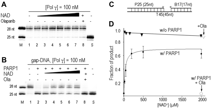
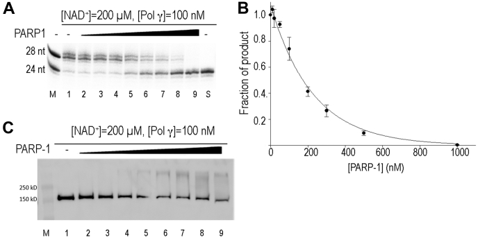
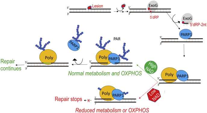
Mitochondrial DNA is located in organelle that house essential metabolic reactions and contains high reactive oxygen species. Therefore, mitochondrial DNA suffers more oxidative damage than its nuclear counterpart. Formation of a repair enzyme complex is beneficial to DNA repair. Recent studies have shown that mitochondrial DNA polymerase (Pol γ) and poly(ADP-ribose) polymerase 1 (PARP1) were found in the same complex along with other mitochondrial DNA repair enzymes, and mitochondrial PARP1 level is correlated with mtDNA integrity. However, the molecular basis for the functional connection between Pol γ and PARP1 has not yet been elucidated because cellular functions of PARP1 in DNA repair are intertwined with metabolism via NAD+ (nicotinamide adenosine dinucleotide), the substrate of PARP1, and a metabolic cofactor. To dissect the direct effect of PARP1 on mtDNA from the secondary perturbation of metabolism, we report here biochemical studies that recapitulated Pol γ PARylation observed in cells and showed that PARP1 regulates Pol γ activity during DNA repair in a metabolic cofactor NAD+ (nicotinamide adenosine dinucleotide)-dependent manner. In the absence of NAD+, PARP1 completely inhibits Pol γ, while increasing NAD+ levels to a physiological concentration that enables Pol γ to resume maximum repair activity. Because cellular NAD+ levels are linked to metabolism and to ATP production via oxidative phosphorylation, our results suggest that mtDNA damage repair is coupled to cellular metabolic state and the integrity of the respiratory chain.

Keywords: ADP-ribosylation; DNA polymerase; DNA repair; DNA synthesis; post-translational modification; protein–DNA interaction; protein–protein interaction; western blot.


Conflict of interest statement

Conflicts of interest The authors declare no conflicts of interest in regards to this manuscript.

Figures

Figure 1
Figure 1
DNA-dependent Pol γ trans-PARylation.Trans-PARylation assays were performed by incubating Pol γ (1 μM) the indicated DNA substrate (0.75 μM) and NAD concentration prior to addition of PARP1 (1 μM). Western blot analysis using antibodies against PolgA was performed (A) and quantified (C) to evaluate trans-PARylation. The membrane was stripped, re-probed with antibodies against PARP1 (B) and quantified (D) to evaluate auto-PARylation. PARP1, Poly(ADP-Ribose) polymerase 1; Pol γ, mitochondrial DNA polymerase.
Figure 2
Figure 2
PARP1 regulates Pol γ gap-filling synthesis.A, Pol γ (100 nM) gap-filling synthesis on 3-nt gapped 32P-DNA substrate (100 nM) in the absence (Lane 1) or presence of NAD+ (20 μM, 50 μM, 200 μM, 500 μM, and 2000 μM, Lanes 2–7) and in the presence of Olaparib (10 μM, Lane 8). Reaction products were separated on 20 or 23% polyacrylamide gels containing 7 M urea and visualized by autoradiography. Lanes M and S denote marker and substrate only lane, respectively. B, same as in A, except 200 nM PARP1 is present. C, substrate for assays containing a 3-nt gap. D, quantification of A and B. PARP1, Poly(ADP-Ribose) polymerase 1; Pol γ, mitochondrial DNA polymerase.
Figure 3
Figure 3
Effects of excess PARP1 on Pol γ gap-filling activity.A, Pol γ (100 nM) gap-filling synthesis on 3-nt gapped 32P-DNA substrate (100 nM) in presence of constant NAD+ (200 μM) and increasing concentration of PARP1 (10 nM, 20 nM, 50 nM, 100 nM, 200 nM, 300 nM, 500 nM, 1000 nM, Lanes 2–9). Reaction products were separated on 20 or 23% polyacrylamide gels containing 7 M urea and visualized by autoradiography. B, western blot of an identical assay (using nonradiolabeled DNA), probed with antibodies against Pol γ showing Pol γ trans-PARylation as a function of PARP1 concentration. C, quantification of A. PARP1, Poly(ADP-Ribose) polymerase 1; Pol γ, mitochondrial DNA polymerase.
Figure 4
Figure 4
PARylation sites identified by LC-MS/MS mapped on a model of PARP1 Pol γ-gap DNA complex composited from crystal structures. PARP1 (blue) is located at the 5’-end of the gap and Pol γ at the 3’-end where Pol γA (orange) is in close contact with PARP1 and Pol γB (light orange) is distal to the DNA gap. The PARylation sites detected are colored red. PARP1, Poly(ADP-Ribose) polymerase 1; Pol γ, mitochondrial DNA polymerase.
Figure 5
Figure 5
PARP1 effect on Pol γ replication.A, Pol γ (100 nM) synthesis on 25/45 nt primer/template DNA (100 nM) in the absence of NAD+ and PARP1 (Lane 1), in the presence of PARP1 (200 nM) but without NAD+ (Lane 2), in the presence of PARP1 (200 nM) and increasing concentrations of NAD+ (20 μM, 50 μM, 200 μM, 500 μM, and 2000 μM, Lanes 3–7), and in the presence of NAD+, PARP1, and Olaparib (10 μM, Lane 8). Reaction products were separated on 20 or 23% polyacrylamide gels containing 7 M urea and visualized by autoradiography. B, quantification of A. PARP1, Poly(ADP-Ribose) polymerase 1; Pol γ, mitochondrial DNA polymerase.
Figure 6
Figure 6
A proposed model for coordinated activity of PARP1 and Pol γ in mitochondrial BER and the association of mtDNA repair and metabolism. OXPHOS stands for oxidative phosphorylation electron transfer chain. BER, base excision repair; PARP1, Poly(ADP-Ribose) polymerase 1; Pol γ, mitochondrial DNA polymerase.

Similar articles

Cited by

References

    1. Brown W.M., George M., Jr., Wilson A.C. Rapid evolution of animal mitochondrial DNA. Proc. Natl. Acad. Sci. U. S. A. 1979;76:1967–1971. - PMC - PubMed
    1. Brown W.M., Prager E.M., Wang A., Wilson A.C. Mitochondrial DNA sequences of primates: Tempo and mode of evolution. J. Mol. Evol. 1982;18:225–239. - PubMed
    1. Almeida K.H., Sobol R.W. A unified view of base excision repair: Lesion-dependent protein complexes regulated by post-translational modification. DNA Repair (Amst) 2007;6:695–711. - PMC - PubMed
    1. Copeland W.C. The mitochondrial DNA polymerase in health and disease. Subcell. Biochem. 2010;50:211–222. - PMC - PubMed
    1. Masson M., Niedergang C., Schreiber V., Muller S., Menissier-de Murcia J., de Murcia G. XRCC1 is specifically associated with poly(ADP-ribose) polymerase and negatively regulates its activity following DNA damage. Mol. Cell. Biol. 1998;18:3563–3571. - PMC - PubMed

Publication types

MeSH terms

LinkOut - more resources