. 2021 Jul;141(7):1664-1674.e7.
doi: 10.1016/j.jid.2020.12.024. Epub 2021 Jan 19.

The Genomic Landscape of Actinic Keratosis

Affiliations

The Genomic Landscape of Actinic Keratosis

Jason Thomson et al. J Invest Dermatol. 2021 Jul.

Abstract

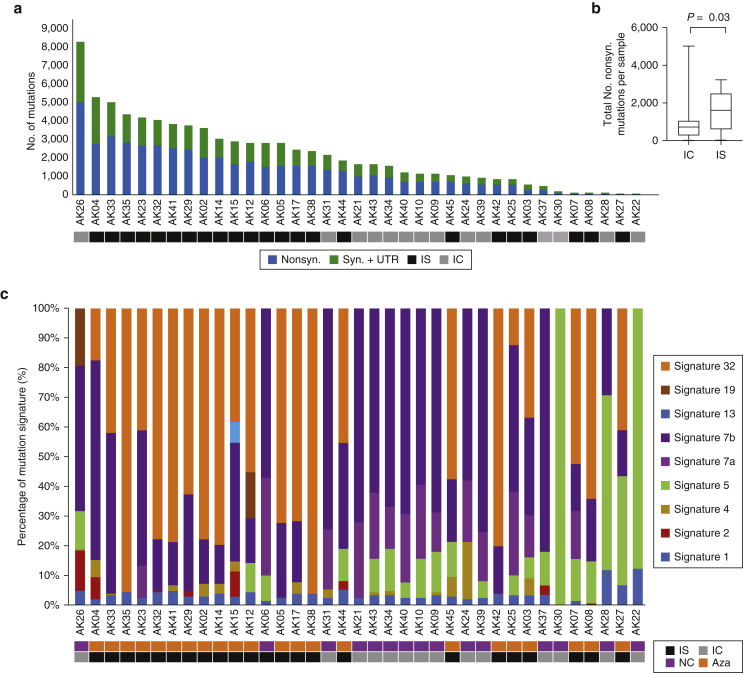
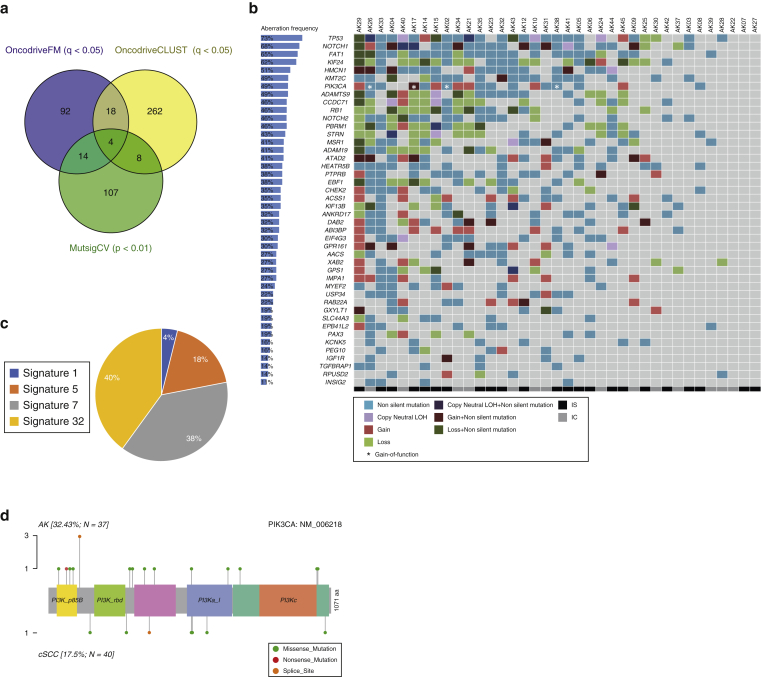
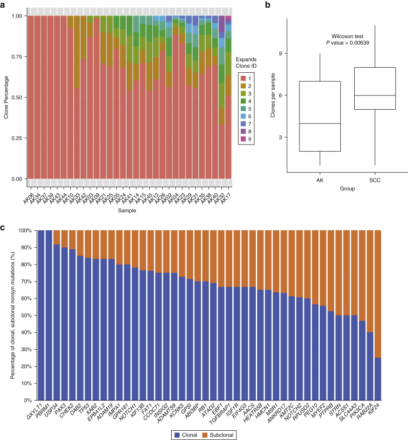
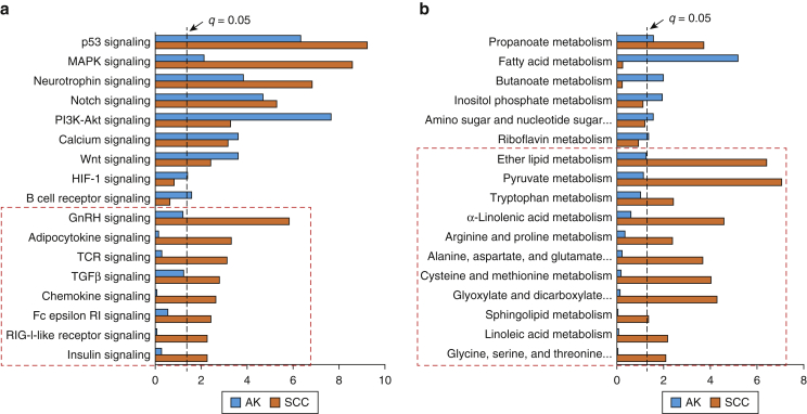
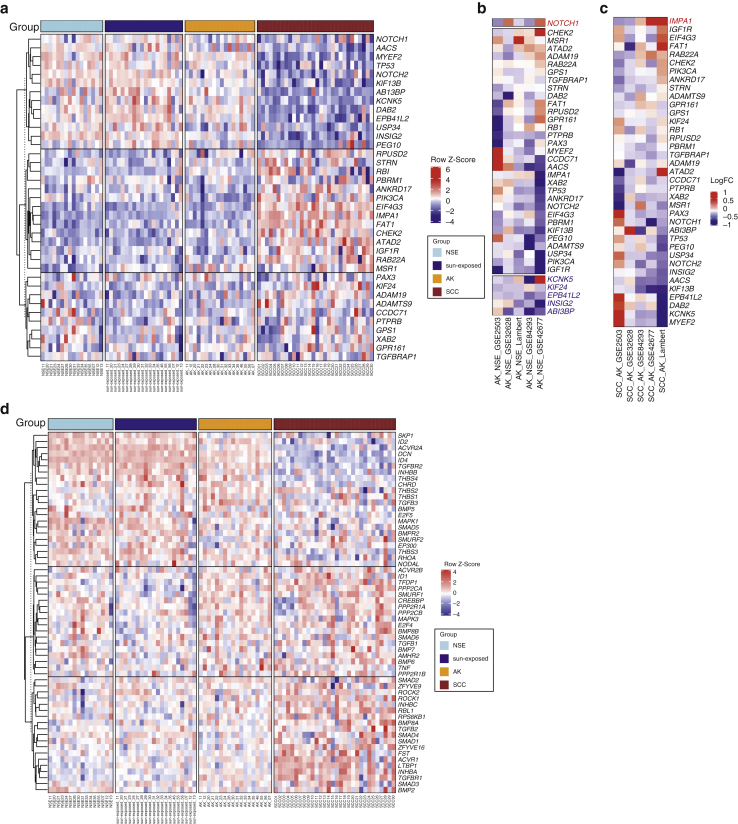
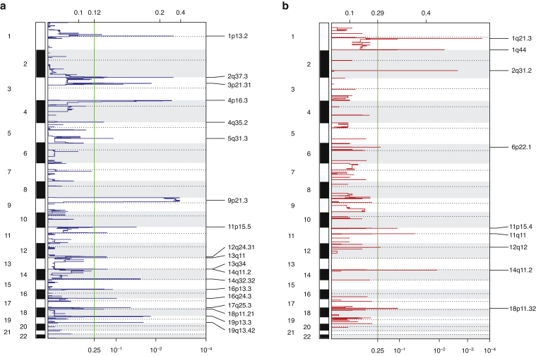
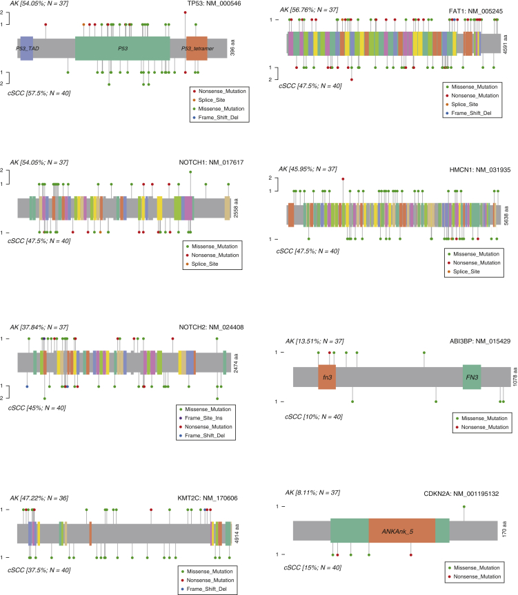
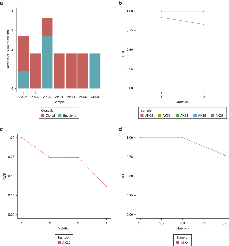
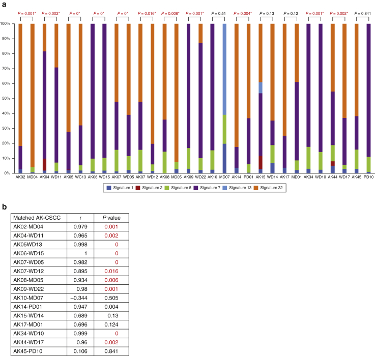
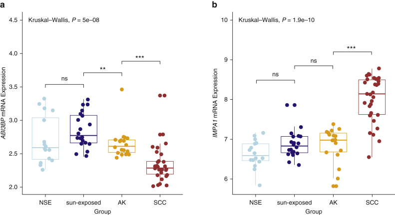
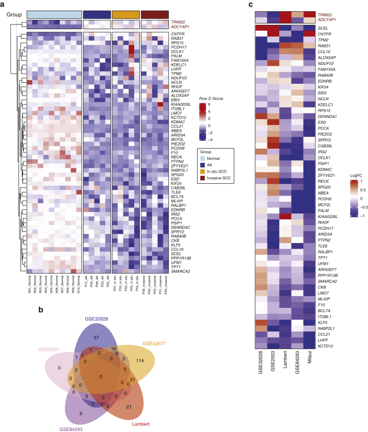
Actinic keratoses (AKs) are lesions of epidermal keratinocyte dysplasia and are precursors for invasive cutaneous squamous cell carcinoma (cSCC). Identifying the specific genomic alterations driving the progression from normal skin to skin with AK to skin with invasive cSCC is challenging because of the massive UVR-induced mutational burden characteristic at all stages of this progression. In this study, we report the largest AK whole-exome sequencing study to date and perform a mutational signature and candidate driver gene analysis on these lesions. We demonstrate in 37 AKs from both immunosuppressed and immunocompetent patients that there are significant similarities between AKs and cSCC in terms of mutational burden, copy number alterations, mutational signatures, and patterns of driver gene mutations. We identify 44 significantly mutated AK driver genes and confirm that these genes are similarly altered in cSCC. We identify azathioprine mutational signature in all AKs from patients exposed to the drug, providing further evidence for its role in keratinocyte carcinogenesis. cSCCs differ from AKs in having higher levels of intrasample heterogeneity. Alterations in signaling pathways also differ, with immune-related signaling and TGFβ signaling significantly more mutated in cSCC. Integrating our findings with independent gene expression datasets confirms that dysregulated TGFβ signaling may represent an important event in AK‒cSCC progression.


Figures

Figure 1
Figure 1
The number of somatic mutations and mutation signatures across 37 AKs. (a) No. of nonsyn., syn., and UTR mutations across the 37 samples. (b) Box and whisker plot comparing the total No. of nonsyn. mutations per AK from IS patients with those from IC patients, showing significantly more mutations in IS patients (Wilcoxon, P = 0.03). (c) Mutation signature compositions across the 37 AK samples. Signature 32 = azathioprine signature, and Signatures 7a/7b = UVR signatures. AK, actinic keratosis; Aza, confirmed azathioprine exposure; IC, immunocompetent; IS, immunosuppressed; NC, no confirmed azathioprine exposure; No., number; syn., synonymous; UTR, untranslated region.
Figure 2
Figure 2
Somatic CNA and LOH events in 37 AKs. OncoPrint of copy-gain, copy-loss, and CN LOH segments with selected known cancer driver genes annotated earlier. AK, actinic keratosis; CN, copy-neutral; CNA, copy number aberration; LOH, loss of heterozygosity.
Figure 3
Figure 3
A total of 44 SMGs identified in the 37 AKs. (a) Venn diagram showing the overlap of numbers of SMGs as assessed by MutsigCV, OncodriveFM, and OncodriveCLUST. (b) Mutation OncoPrint of the 44 SMGs identified by at least two of the three methods, with CNA integrated and overall alteration frequency indicated with percentage bars. (c) Pie chart of the mutational signature contribution to 44 SMG mutations. (d) Lollipop plot of PIK3CA showing the distribution of mutation in AK and cSCC cohorts. aa, amino acid; AK, actinic keratosis; CNA, copy number aberration; cSCC, cutaneous squamous cell carcinoma; IC, immunocompetent; IS, immunosuppressed; LOH, loss of heterozygosity; PI3K, phosphoinositide 3-kinase; SMG, significantly mutated gene.
Figure 4
Figure 4
Clonality analysis in AK. (a) EXPANDS clonality analysis showing clones and their percentages per sample. (b) Box and whisker plot comparing clone sizes between AK and cSCC. P = 0.00639. (c) Clonality analysis of 44 SMGs identifying clonal and subclonal nonsyn mutations using EXPANDS. AK, actinic keratoses; cSCC, cutaneous squamous cell carcinoma; EXPANDS, Expanding Ploidy and Allele-frequency on Nested Subpopulations; ID, identity; SCC, squamous cell carcinoma; SMG, significantly mutated gene; syn, synonymous.
Figure 5
Figure 5
Comparison of significantly mutated KEGG pathways between AK and cSCC. (a) Significantly mutated signaling pathways derived from OncodriveFM. (b) Significantly mutated metabolic pathways. Pathways and biological processes in the bar charts were sorted with terms significant (cutoff q = 0.05) in both groups at the top and terms significant in cSCC only at the bottom (indicated with red dashed box). AK, actinic keratosis; Akt, protein kinase B; cSCC, cutaneous squamous cell carcinoma; KEGG, Kyoto Encyclopedia of Genes and Genomes; PI3K, phosphoinositide 3-kinase; SCC, squamous cell carcinoma.
Figure 6
Figure 6
Expression profiles of SMGs in the five independent gene expression datasets. (a) Expression heatmap of 37 SMGs across NSE skin, sun-exposed skin, AK, and cSCC samples from the Lambert dataset. LogFC of pairwise comparisons of (b) AK versus NSE and (c) SCC versus AK across the five datasets (Lambert, GSE42677, GSE84293, GSE2503, and GSE32628). Normal skin: NSE skin, sun-exposed skin, AK, and SCC‒cSCC. Significant genes with the same direction in 2 of 5 datasets were highlighted in blue (downregulated) or in red (upregulated). Within b and c, the blue color indicates the downregulation in AK or SCC compared with the downregulation in NSE or AK, respectively, and the red color indicates the upregulation in AK or SCC relative to that in NSE or AK, respectively. (d) Expression heatmap of TGFβ signaling genes that were expressed across NSE skin, sun-exposed skin, AK, and cSCC samples from the Lambert dataset. AK, actinic keratosis; cSCC, cutaneous squamous cell carcinoma; LogFC, Log2 fold change; NSE, non–sun-exposed; SCC, squamous cell carcinoma; SMG, significantly mutated gene.
Supplementary Figure S1
Significantly (a) deleted and (b) gained regions across the 33 AK samples based on GISTIC2.0. Four samples were excluded from the analysis because they have too few CNA segments. AK, actinic keratosis; CNA, copy number aberration.
Supplementary Figure S2
Lollipop plots of key AK and cSCC driver genes showing the type and distribution patterns of nonsynonymous mutations between AK and cSCC. aa, amino acid; AK, actinic keratosis; cSCC, cutaneous squamous cell carcinoma; Del, deletion; Ins, insertion.
Supplementary Figure S3
Boxplots of CCFs of synonymous and nonsynonymous mutations of SMGs between AK and cSCC, highlighting that two genes had more clonal nonsynonymous mutations in AK than in (a) CACNA1C and (b) KCNK5, using synonymous mutations as the negative control. AK, actinic keratosis; CCF, cancer cell fraction; SMG, significantly mutated gene; SCC, squamous cell carcinoma.
Supplementary Figure S4
TP53 clonality analysis. (a) Bar chart of the number of clonal and subclonal TP53 mutations in selected AK samples with >1 nonsynonymous TP53 mutation. (b–d) Line graphs of CCF of TP53 nonsynonymous mutations for (b) AK05, AK33, AK34, AK38, (c) AK32, and (d) AK04. AK, actinic keratosis; CCF, cancer cell fraction.
Supplementary Figure S5
Correlation of mutation signatures between matched AK and cSCC pairs. (a) Mutation signature compositions in matched AK and cSCC pairs. Two pairs (AK03‒MD03 and AK29‒WD08) were not included because the cSCC did not have mutational signature data. P-values from Pearson correlation statistic. Signature 32 = azathioprine signature, and signature 7 = UVR signature. (b) Table of Pearson correlation analysis. AK, actinic keratosis; cSCC, cutaneous squamous cell carcinoma; MD, moderately differentiated; WD, well-differentiated.
Supplementary Figure S6
Box and whisker plot comparing (a) ABI3BP with (b) IMPA1 mRNA expression across NSE skin, sun-exposed skin, AK, and cSCC, depicting the statistics for between-group comparisons. ns and significant at ∗∗P < 0.01 and ∗∗∗P< 0.001. AK, actinic keratosis; cSCC, cutaneous squamous cell carcinoma; ns, not significant; NSE, non–sun-exposed; SCC, squamous cell carcinoma.
Supplementary Figure S7
Expression profiles of genes significantly gained (red) or deleted (black) in AK. (a) Expression heatmap of significantly gained, upregulated genes (n = 2, red) and deleted, downregulated genes (n = 52, black) in the AK versus those in the normal skin that were also expressed across normal skin, AK, and in situ and invasive cSCC samples from the GSE42677. (b) Venn diagram of overlap across the five gene expression datasets of significantly deleted and downregulated genes in AK versus those in the normal skin. (c) LogFC of significant genes with the same direction in 2 of 5 datasets in pairwise comparisons of AK with normal skin across the five datasets (Lambert, GSE42677, GSE84293, GSE2503, and GSE32628). The blue color indicates the downregulation in AK compared with that in the normal skin, and the red color indicates the upregulation in AK relative to that in the normal skin. AK, actinic keratosis; cSCC, cutaneous squamous cell carcinoma; LogFC, Log2 fold change.

Comment in

References

    1. Albibas A.A., Rose-Zerilli M.J.J., Lai C., Pengelly R.J., Lockett G.A., Theaker J. Subclonal evolution of cancer-related gene mutations in p53 immunopositive patches in human skin. J Invest Dermatol. 2018;138:189–198. - PubMed
    1. Alexandrov L.B., Kim J., Haradhvala N.J., Huang M.N., Tian Ng A.W.T., Wu Y. The repertoire of mutational signatures in human cancer. Nature. 2020;578:94–101. - PMC - PubMed
    1. Alexandrov L.B., Nik-Zainal S., Wedge D.C., Campbell P.J., Stratton M.R. Deciphering signatures of mutational processes operative in human cancer. Cell Rep. 2013;3:246–259. - PMC - PubMed
    1. Andor N., Harness J.V., Müller S., Mewes H.W., Petritsch C. EXPANDS: expanding ploidy and allele frequency on nested subpopulations. Bioinformatics. 2014;30:50–60. - PMC - PubMed
    1. Ashton K.J., Weinstein S.R., Maguire D.J., Griffiths L.R. Chromosomal aberrations in squamous cell carcinoma and solar keratoses revealed by comparative genomic hybridization. Arch Dermatol. 2003;139:876–882. - PubMed

Publication types

MeSH terms

Substances