. 2021;11(5):1369-1385.
doi: 10.1016/j.jcmgh.2021.01.007. Epub 2021 Jan 19.

The Ubiquitin E3 Ligase TRIM21 Promotes Hepatocarcinogenesis by Suppressing the p62-Keap1-Nrf2 Antioxidant Pathway

Affiliations

The Ubiquitin E3 Ligase TRIM21 Promotes Hepatocarcinogenesis by Suppressing the p62-Keap1-Nrf2 Antioxidant Pathway

Fang Wang et al. Cell Mol Gastroenterol Hepatol. 2021.

Abstract

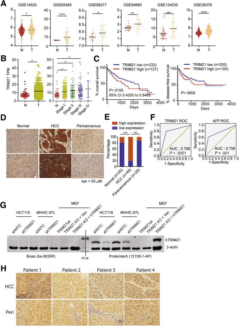
Background and aims: TRIM21 is a ubiquitin E3 ligase that is implicated in numerous biological processes including immune response, cell metabolism, redox homeostasis, and cancer development. We recently reported that TRIM21 can negatively regulate the p62-Keap1-Nrf2 antioxidant pathway by ubiquitylating p62 and prevents its oligomerization and protein sequestration function. As redox homeostasis plays a pivotal role in many cancers including liver cancer, we sought to determine the role of TRIM21 in hepatocarcinogenesis.

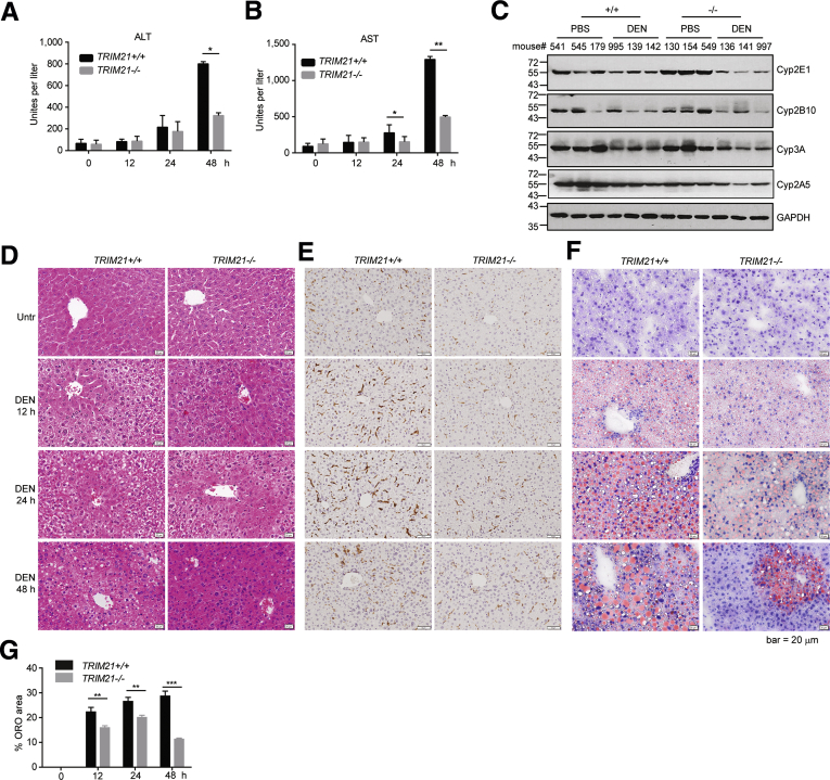
Methods: We examined the correlation between TRIM21 expression and the disease using publicly available data sets and 49 cases of HCC clinical samples. We used TRIM21 genetic knockout mice to determine how TRIM21 ablation impact HCC induced by the carcinogen DEN plus phenobarbital (PB). We explored the mechanism that loss of TRIM21 protects cells from DEN-induced oxidative damage and cell death.

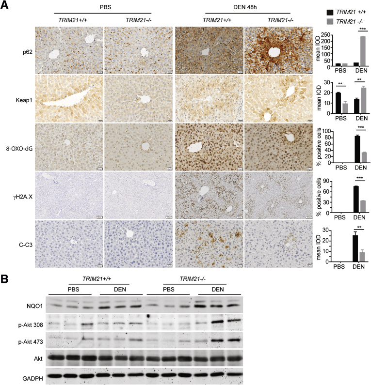
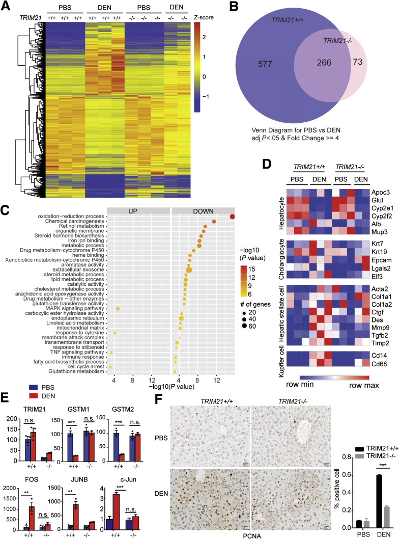
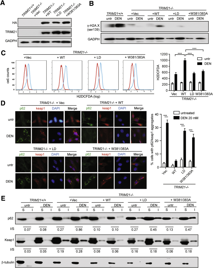
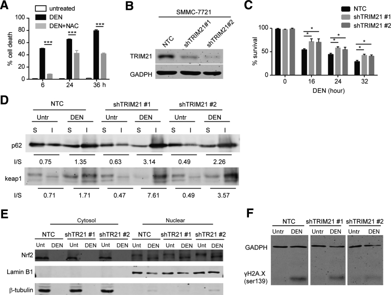
Results: There is a positive correlation between TRIM21 expression and HCC. Consistently, TRIM21-knockout mice are resistant to DEN-induced hepatocarcinogenesis. This is accompanied by decreased cell death and tissue damage upon DEN treatment, hence reduced hepatic tissue repair response and compensatory proliferation. Cells deficient in TRIM21 display enhanced p62 sequestration of Keap1 and are protected from DEN-induced ROS induction and cell death. Reconstitution of wild-type but not the E3 ligase-dead and the p62 binding-deficient mutant TRIM21 impedes the protection from DEN-induced oxidative damage and cell death in TRIM21-deficient cells.

Conclusions: Increased TRIM21 expression is associated with human HCC. Genetic ablation of TRIM21 leads to protection against oxidative hepatic damage and decreased hepatocarcinogenesis, suggesting TRIM21 as a preventive and therapeutic target.

Keywords: Diethylnitrosamine; Hepatocellular Carcinoma; Nrf2; TRIM21; p62.


Figures

Figure 1
Figure 1
Increased TRIM21 expression is associated with HCC. (A) In the Gene Expression Omnibus Series, 6 studies showed elevated TRIM21 expression in HCC compared with normal liver tissues. (B) TCGA shows increased TRIM21 TPM in liver cancer comparing with normal tissues. (C)TRIM21 high expression indicates poor prognosis with decreasing overall survival according to TCGA data analysis, while the disease-free survival is trending lower yet not significant. (D) Liver tissue sections were stained for TRIM21 by immunohistochemistry (IHC). Representative TRIM21 staining on normal liver tissues from peripheral hepatic hemangioma (left panel; n = 20), on tumor tissues from HCC (middle panel; n = 49), and on paracancerous tissues 2 cm away from the HCC (right panel; n = 29). (E) TRIM21 expression is increased in HCC tissues compared with the paracancerous tissues and normal liver tissues. Relative intensity of staining in each section was assessed blindly and assigned a value ranging from 0 to 4 according to intensity of staining: score 0, negative staining, no positive cells; score 1, weak staining, the ratio of positive cells to total cells ≤25%; score 2, moderately positive, the ratio of positive cells to total cells ≤50%; score 3, moderately positive, the ratio of positive cells to total cells ≤75%; score 4, strongly positive, the ratio of positive cells to total cells>75%. Low expression defines the score from 0 to 2 and high expression is the score 3–4. (F) Receiver-operating characteristic (ROC) curves of the ability of TRIM21 or AFP high expression to the diagnosis of HCC. (G) TRIM21 was silenced in HCT116 and MHHC-97L human cells, or reconstituted with vector or HA-hTRIM21 in TRIM21–/– MEFs. Cells were lysed in RIPA buffer (1% SDS) and subjected to WB. The TRIM21 antibody obtained from Bioss (bs-0635R) or Proteintech (12018-1-AP) was used to detect human TRIM21. Note that the Bioss antibody fails to recognize human TRIM21, and neither antibody recognizes murine TRIM21. (H) TRIM21 antibody (Bioss bs-0635R) was used for IHC on HCC tumor tissues and paracancerous tissues collected 2 cm away from the HCC. Note that the Bioss antibody fails to detect difference in IHC staining in paracancerous and HCC tissues. ∗P < .05. ∗∗P < .01. ∗∗∗P < .001.
Figure 2
Figure 2
TRIM21 ablation leads to decreased HCC induced by DEN/PB. (A) TRIM21+/+ (n = 8), TRIM21+/– (n = 15), and TRIM21–/– (n = 14) 14-day-old male mice were intraperitoneal injected with DEN (5 mg/kg), then fed with 0.05% PB in drinking water 7 days later. Livers from DEN/PB-treated mice were collected after 10 months. Representative images are shown. (B) Liver weight and body weight were measured 10 months after DEN/PB treatment. Ratio of liver weight to body 8 was plotted. (C) Total tumor number was counted and plotted. Each dot represents 1 mouse. (D) Tumors with >0.3 cm diameter was counted and plotted. (E) Liver tissue sections were processed for hematoxylin and eosin staining. TRIM21+/+ (left panel), TRIM21+/– (middle panel), and TRIM21–/– (right panel) liver tissue showed solid type hepatocellular carcinoma, with poorly differentiated (including tumor giant cell), moderately differentiated, and well differentiated, respectively. (F) Liver tissues sections were stained for HSP70 and PCNA. PCNA-positive cells were counted and the means of 3 randomly selected fields are shown. (G) Liver tissue sections were processed for Sirius Red staining and α-SMA/DAPI staining by immunofluorescence. Representative images are shown. ∗P < .05. ∗∗P < .01. ∗∗∗P < .001. IOD, integrated optical density.
Figure 3
Figure 3
TRIM21–/–livers are protected from DEN-induced liver damage. (A, B) Eight-week-old TRIM21+/+ and TRIM21–/– male mice were treated with 200-mg/kg DEN via intraperitoneal injection. Cardiac blood was collected. (A) Alanine aminotransferase and (B) aspartate aminotransferase were measured before and after 12, 24, and 48 hours treatment. (C) Liver tissues (control and 48 hours DEN treatment) were lysed in RIPA buffer and subjected to WB for enzymes related to DEN metabolism. (D) Hematoxylin and eosin staining of formalin-fixed, paraffin-embedded liver sections. (E) F-4/80 staining indicates more profound monocyte infiltration in WT livers upon DEN treatment. (F) Oil Red O (ORO) staining of frozen liver sections with hematoxylin counterstaining. Red indicates lipid, purple indicates nuclei. Representative images are shown. (G) Quantitation of the area occupied by lipid inclusions within the frozen liver sections after staining of sections with ORO. Data are presented as mean ± SEM of 3 replicates. ∗∗P < .01. ∗∗∗P < .001.
Figure 4
Figure 4
TRIM21–/–livers are protected from DEN-induced liver damage. (A) Eight-week-old TRIM21+/+ and TRIM21–/– mice were treated with 200-mg/kg DEN via intraperitoneal injection. Livers were collected 48 hours later and processed for IHC analysis. Mean integrated optical density was determined for p62, Keap1, and cleaved caspase-3 (C-C3) because they are predominantly localized in the cytosol. Cells with positive nuclear staining of 8-oxo-dG and γH2A.X staining were counted and indicated as the percentage to total cells. Representative images are shown. Data are presented as mean ± SEM of 3 replicates. (B) Liver tissues were lysed in RIPA buffer and subjected to WB for indicated proteins. ∗∗P < .01. ∗∗∗P < .001.
Figure 5
Figure 5
DEN-induced hepatocyte proliferation is suppressed in TRIM21–/–liver. (A) Eight-week-old TRIM21+/+ and TRIM21–/– male mice were treated with PBS or 200-mg/kg DEN by intraperitoneal injection. Liver tissues were collected after 48 hours for RNA-seq. Hierarchical clustering and heatmap illustration of differentially expressed genes under indicated conditions. Color key indicates Z-scores after normalization. (B) Venn diagram showing overlap of genes differentially expressed in PBS-treated vs DEN-treated TRIM21+/+ and TRIM21–/– mice. Analysis was restricted to genes, which showed a 4-fold difference and yielded P < .05. (C) Dot plots showing the enriched terms from Gene Ontology analysis using differentially expressed genes identified from DEN vs PBS treatment in TRIM21 WT mice (the 577 unique genes altered by >4-fold in TRIM21-WT mice as shown in B). Note that genes involved in redox regulation, chemical carcinogenesis, and retinol metabolism are identified. (D) Heatmap shows that DEN-induced expressions of hepatic stellate cell and Kupffer cell markers were markedly decreased in TRIM21 KO livers, while no obvious difference was detected in hepatocyte markers. (E) Quantitative reverse-transcription PCR for the DEN-treated TRIM21 WT and KO liver samples, showing decreased antioxidant response and increased proliferation signals DEN-treated WT mice (n = 3 per group). (F) IHC analysis of PCNA expression in livers of PBS- and DEN-treated TRIM21 WT and KO mice. PCNA-nuclear positive cells were counted and the ratio of positive cells was calculated. Representative images are shown. Data are presented as means ± SEM of 3 mice in each group. ∗∗∗P < .001. FPKM, fragments per kilobase of transcript per million; n.s., nonsignificant.
Figure 6
Figure 6
TRIM21 silencing protects cells from DEN-induced oxidative damage.TRIM21–/– MEFs reconstituted with vector, HA-TRIM21 WT, HA-TRIM21 LD, and HA-TRIM21 W381/383A mutant. (A) Cells were lysed in RIPA buffer with 1% SDS and subjected to WB. (B–E) Cells were treated with DEN (20 mM) for 6 hours. (B) Cells were harvested and probed for indicated proteins by WB. (C) Cells were stained with H2DCFDA and analyzed by flow cytometry. Quantification of relative H2DCFDA intensity (geometry mean of 3 repeats) is shown on the right. ∗∗∗P < .001. (D) Cells were subjected to IF with Keap1 and p62 antibodies and observed under deconvolution microscope. Cells with p62/Keap1 aggregates were counted blindly. Data shown are the averages plus SD of at least 3 countings with over 200 cells. ∗∗∗P < .001. (E) Cells were separated into Triton X-100 (1%) soluble and insoluble fractions, and 30-μg soluble proteins and corresponding volume of insoluble proteins were used for WB. The numbers below Western blot panels indicate the ratios of absolute levels of insoluble and soluble proteins, respectively.
Figure 7
Figure 7
Silencing of TRIM21 protects cells from DEN-induced genotoxic damage and cell death. (A) SMMC-7721 cells were treated with DEN (50 mM) alone or together with N-acetylcysteine (5 mM) for indicated time. Cell viability was analyzed by PI exclusion. ∗∗∗P < .001. (B) TRIM21 was silenced in SMMC-7721 cells using 2 independent short hairpin RNAs. Cells was lysed in RIPA buffer with 1% SDS and subjected to WB. (C) SMMC-7721 with nontargeted control, shTRIM21 #1 and shTRIM21 #2 were treated with DEN (50 mM) for indicated periods of time. Cell viability was determined by trypan blue exclusion. Shown are the mean plus SD of 3 countings. ∗P < .05. (D) SMMC-7721 with nontargeted control, shTRIM21 #1, and shTRIM21 #2 were treated with DEN (50 mM) for 6 hours. Cells were lysed in immunoprecipitation lysis buffer containing 1% Triton X-100. The insoluble fraction was dissolved in RIPA buffer containing 1% SDS. Both Triton X-100 soluble and insoluble fractions were subjected to WB, and 30-μg soluble proteins and corresponding volume of insoluble proteins were used for WB. The numbers below Western blot panels indicate the ratios of absolute levels of insoluble and soluble proteins, respectively. (E) SMMC-7721 cells were treated with DEN (50 mM) for 6 hours. Cells were separated into cytoplasmic and nuclear fractions, and 20-μg cytosolic proteins and corresponding volume of nuclear fractions were used for WB. (F) SMMC-7721 cells were treated with DEN (50 mM) for 6 hours. Cells was lysed in RIPA buffer (1% SDS) and subjected to WB.

Similar articles

Cited by

References

    1. Choi J. Oxidative stress, endogenous antioxidants, alcohol, and hepatitis C: pathogenic interactions and therapeutic considerations. Free Radic Biol Med. 2012;52:1135–1150. - PubMed
    1. Gambino R., Musso G., Cassader M. Redox balance in the pathogenesis of nonalcoholic fatty liver disease: mechanisms and therapeutic opportunities. Antioxid Redox Signal. 2011;15:1325–1365. - PubMed
    1. Ivanov A.V., Valuev-Elliston V.T., Tyurina D.A., Ivanova O.N., Kochetkov S.N., Bartosch B., Isaguliants M.G. Oxidative stress, a trigger of hepatitis C and B virus-induced liver carcinogenesis. Oncotarget. 2017;8:3895–3932. - PMC - PubMed
    1. Dodson M., de la Vega M.R., Cholanians A.B., Schmidlin C.J., Chapman E., Zhang D.D. Modulating NRF2 in disease: timing is everything. Annu Rev Pharmacol Toxicol. 2019;59:555–575. - PMC - PubMed
    1. Komatsu M., Kurokawa H., Waguri S., Taguchi K., Kobayashi A., Ichimura Y., Sou Y.S., Ueno I., Sakamoto A., Tong K.I., Kim M., Nishito Y., Iemura S., Natsume T., Ueno T., Kominami E., Motohashi H., Tanaka K., Yamamoto M. The selective autophagy substrate p62 activates the stress responsive transcription factor Nrf2 through inactivation of Keap1. Nat Cell Biol. 2010;12:213–223. - PubMed

Publication types

MeSH terms