. 2021 Jan 22;17(1):45.
doi: 10.1186/s12917-020-02736-2.

A case of a complete atrioventricular canal defect in a ferret

Affiliations

A case of a complete atrioventricular canal defect in a ferret

Carlos F Agudelo et al. BMC Vet Res. .

Abstract

Background: Atrioventricular canal defect is a rare congenital disorder of the heart and describes the presence of an atrial septal defect, a variable presentation of ventricular septal alterations including ventricular septal defect malformations in the mitral and tricuspid valves. The defect has been described in human beings, dogs, cats, pigs, and horses.

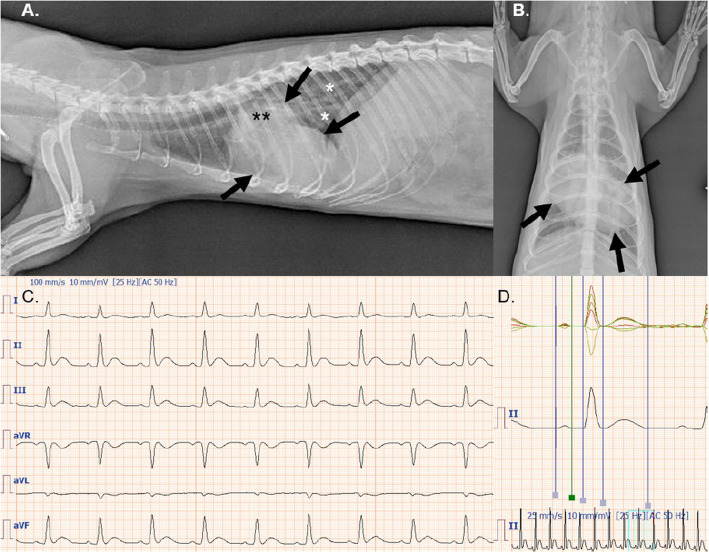
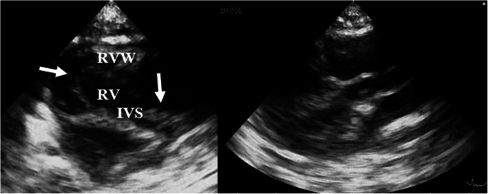
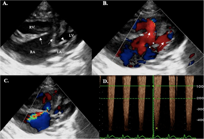
Case presentation: This paper describes the case of a complete atrioventricular canal defect in a four-year-old intact male pet ferret (Mustela putorius furo), which was presented due to posterior weakness, ataxia, and decreased appetite. A loud systolic murmur, dyspnea, and hind limb paraparesis were detected during the clinical examination. Thoracic radiographs showed generalized cardiomegaly and lung edema. ECG showed sinus rhythm with prolonged P waves and QRS complexes. Echocardiography showed a large atrial septal defect, atrioventricular dysplasia, and a ventricular septal defect. Palliative treatment with oxygen, furosemide, spironolactone, enalapril, diltiazem, and supportive care was chosen as the therapy of choice. The ferret recovered gradually during hospitalization. A follow-up examination at three and six months showed stabilization of cardiac function.

Conclusions: To the authors knowledge, this is the first time an atrioventricular canal defect has been described in a pet ferret.

Keywords: Atrioventricular canal; Echocardiography; Endocardial cushion defect; Heart failure; Pet ferret.


Conflict of interest statement

The authors declare that they have no competing interests.

Figures

Fig. 1
Fig. 1
a Left laterolateral projection shows generalized cardiomegaly (vertebral heart score = 8) namely both ventricles and the left atrium (black arrows) with dorsal displacement of the trachea. There is also venous congestion (black asterisks) and bronchial lung pattern presence on the caudal lung lobes (white asterisks). b The dorsoventral view also demonstrates marked biventricular and left atrial enlargement. c Sinus rhythm (regular RR intervals) at a rate of 200–210 bpm. 100 mm / s and 1 cm = 10 mV. d Digital simultaneous superimposition of limb leads enhancing the measurement of diferent waves and intervals. There is a prolongation of the P wave suggesting left atrial (0.04 s; normal 0.01–0.03 s) and left ventricular enlargement (0.055 s; normal 0.02–0.05 s) [19]
Fig. 2
Fig. 2
Left: Right parasternal short axis view displaying right ventricular hypertrophy and IVS at the level of papillary muscles. Flattening of the interventricular septum is present. There is present tricuspid valve deformation showing abnormal inserted portions of the valve leaflets (white arrows). Right: Mildly anteriorly displaced aortic valve with elongated LVOT (goose neck). Key: IVS: interventricular septum; RV: right ventricle; RVW: right ventricular wall
Fig. 3
Fig. 3
Simultaneous B-mode, CFM and CW imaging in a modified left apical 5-chamber view. a Marked right atrial enlargement can be noticed with a markedly thickened tricuspidal leaflets (white arrows) and absence of the atrial septum. Anterior mitral leaflet (white arrowhead). b AV inflow shows common filling of both ventricles (white arrows) and defects position (asteriscs). c CFM exam shows a large tricuspid regurgitation that reach portions of the left atrium. d CW Doppler. The cursor is located across the tricuspid regurgitation at the beginning of the systole and shows a velocity of 4.39 m/s (82 mmHg). Key: LA: left atrium; RA: right atrium, RV: right ventricle

References

    1. Schrope DP. Atrioventricular septal defects: natural history, echocardiographic, electrocardiographic, and radiographic findings in 26 cats. J Vet Cardiol. 2013;15:233–42. doi: 10.1016/j.jvc.2013.06.004. - DOI - PubMed
    1. Saponaro V, Staffieri F, Franchini D, Crovace A. Complete atrioventricular canal in a dog. J Vet Cardiol. 2010;12:135–40. doi: 10.1016/j.jvc.2010.04.003. - DOI - PubMed
    1. Lee SG, Nam SJ, Moon HS, Hyun CB. Partial atrioventricular canal defect in a maltese dog. J Vet Clin. 2008;25:195–99.
    1. Amberger CN, Boujon CE, Amberger C, Boujon C. Atrioventricular canal defect (incomplete form of endocardial cushion defect) and ostium secundum type interatrial septal defect in a dog. J Vet Cardiol. 2002;4:37–41. doi: 10.1016/S1760-2734(06)70037-6. - DOI - PubMed
    1. Hsu FS, Du SJ. Congenital heart diseases in swine. Vet Pathol. 1982;19:676–86. doi: 10.1177/030098588201900613. - DOI - PubMed

MeSH terms

Supplementary concepts

LinkOut - more resources