. 2021 Jan 22;16(1):3.
doi: 10.1186/s13024-021-00423-w.

Methods to investigate intrathecal adaptive immunity in neurodegeneration

Affiliations

Methods to investigate intrathecal adaptive immunity in neurodegeneration

Hamilton Oh et al. Mol Neurodegener. .

Abstract

Background: Cerebrospinal fluid (CSF) provides basic mechanical and immunological protection to the brain. Historically, analysis of CSF has focused on protein changes, yet recent studies have shed light on cellular alterations. Evidence now exists for involvement of intrathecal T cells in the pathobiology of neurodegenerative diseases. However, a standardized method for long-term preservation of CSF immune cells is lacking. Further, the functional role of CSF T cells and their cognate antigens in neurodegenerative diseases are largely unknown.

Results: We present a method for long-term cryopreservation of CSF immune cells for downstream single cell RNA and T cell receptor sequencing (scRNA-TCRseq) analysis. We observe preservation of CSF immune cells, consisting primarily of memory CD4+ and CD8+ T cells. We then utilize unbiased bioinformatics approaches to quantify and visualize TCR sequence similarity within and between disease groups. By this method, we identify clusters of disease-associated, antigen-specific TCRs from clonally expanded CSF T cells of patients with neurodegenerative diseases.

Conclusions: Here, we provide a standardized approach for long-term storage of CSF immune cells. Additionally, we present unbiased bioinformatic approaches that will facilitate the discovery of target antigens of clonally expanded T cells in neurodegenerative diseases. These novel methods will help improve our understanding of adaptive immunity in the central nervous system.

Keywords: Adaptive immunity; Antigen; CSF; Cerebrospinal fluid cells; Intrathecal cells; Neurodegeneration; T cell receptor (TCR); T cells.


Conflict of interest statement

The authors declare that they have no competing interests.

Figures

Fig. 1
Fig. 1
Cryopreservation of human cerebrospinal fluid cells retains cellular viability. a Workflow for cryopreservation of CSF cells for scRNA-TCRseq. b Gating strategy for sorting live cryopreserved cells by flow cytometry. c Quantification of live cell count (live singlets) from flow cytometry sorting following cell thawing. Mean ± s.e.m. d Quantification of viability (percentage of live singlets out of all singlets) from sorting by flow cytometry. Mean ± s.e.m. e Linear regression of live cell count post thaw versus length of storage. f Quantification of the number of CSF cells captured for sequencing
Fig. 2
Fig. 2
scRNA-TCRseq of human cerebrospinal fluid reveals clonally expanded CD8+ T cells. a Seurat dimensionality reduction and clustering of 22 CSF samples that passed quality control displayed on tSNE (GNLY: Granulysin; DCs: Dendritic Cells). b Heatmap of cluster marker genes used to annotate clusters. c Cells with detected TCR displayed on tSNE. d CD3D expression displayed on tSNE. e Quantification of average cell type distribution based on Seurat clustering. f Clones – cells with TCR sequences shared with other cells – displayed on tSNE. Only cells with detected TCRs are included. g Clones of different sizes displayed on tSNE. Only cells with detected TCRs are included; lower right heatmap shows significant differentially expressed genes between clone size bins. h Quantification of number of T cell types per clone size. Only cells with detected TCR are included. GNLY+CD8+ T Cells and CD8+ T cells were combined as CD8+ T cells. i Quantification of % T cell types per clone size
Fig. 3
Fig. 3
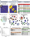
Analysis of T cell receptor sequence similarity within and between neurodegenerative disease groups. a Workflow for quantifying TCRαβ similarity. Clonal TCRs with unambiguous CDR3a and CDR3b sequences were retained for analysis. L-sim values were then computed between all possible combinations of TCRαβ sequences. b At left: heatmap highlighting TCR combinations with L-sim score > 0.8. At right: inset of heatmap shows three clusters of similar TCRs. Color bar represents L-sim score. c Metadata of clustered TCRs shown in b). Note the similarity of TCRs within disease groups. d Three clusters of similar TCRs identified in c) highlighted on tSNE. Clusters of TCRs overlap with CD8+ T cells. e TCR network displaying connections between samples with similar sequences (L-sim > 0.8) identified in b). Each node is a unique patient sample with each small circle sprouting off a node representing a unique clonal TCR. Nodes are colored by disease group; lower right heatmap shows number of similar TCRs between unique samples per disease group. f TCR network (L-sim > 0.9). g Quantification of shared motifs present in TCRβ sequences. Clonal TCRs with unambiguous CDR3b sequences were retained for analysis. The first and last two amino acids were removed from the analysis, since these regions are highly conserved. h Table of motifs of length 9. The SSLGQAYEQ motif is highlighted and metadata corresponding to patient samples is shown. i Upper: table of pathogens specific to SSLGQAYEQ motif based on search of the public McPAS-TCR database. Lower: table of SSLGQAYEQ motif-containing T cell types based on public McPAS-TCR database search

References

    1. Han S, Lin YC, Wu T, Salgado AD, Mexhitaj I, Wuest SC, et al. Comprehensive immunophenotyping of cerebrospinal fluid cells in patients with neuroimmunological diseases. J Immunol. 2014;192(6):2551–2563. doi: 10.4049/jimmunol.1302884. - DOI - PMC - PubMed
    1. Perrin RJ, Fagan AM, Holtzman DM. Multimodal techniques for diagnosis and prognosis of Alzheimer's disease. Nature. 2009;461(7266):916–922. doi: 10.1038/nature08538. - DOI - PMC - PubMed
    1. Blennow K, Zetterberg H. The past and the future of Alzheimer's disease fluid biomarkers. J Alzheimers Dis. 2018;62(3):1125–1140. doi: 10.3233/JAD-170773. - DOI - PMC - PubMed
    1. Ransohoff RM, Engelhardt B. The anatomical and cellular basis of immune surveillance in the central nervous system. Nat Rev Immunol. 2012;12(9):623–635. doi: 10.1038/nri3265. - DOI - PubMed
    1. Schafflick D, Xu CA, Hartlehnert M, Cole M, Schulte-Mecklenbeck A, Lautwein T, et al. Integrated single cell analysis of blood and cerebrospinal fluid leukocytes in multiple sclerosis. Nat Commun. 2020;11(1):247. doi: 10.1038/s41467-019-14118-w. - DOI - PMC - PubMed

Publication types