. 2021 Jan 22;52(1):12.
doi: 10.1186/s13567-020-00882-x.

Activation of regulated cell death in the lung of piglets infected with virulent PRRSV-1 Lena strain occurs earlier and mediated by cleaved Caspase-8

Affiliations

Activation of regulated cell death in the lung of piglets infected with virulent PRRSV-1 Lena strain occurs earlier and mediated by cleaved Caspase-8

Jose María Sánchez-Carvajal et al. Vet Res. .

Abstract

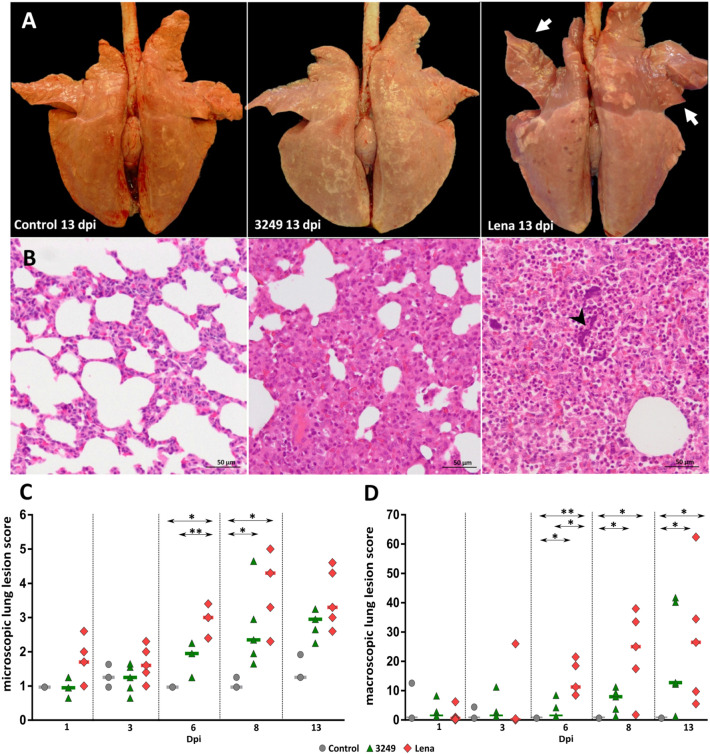
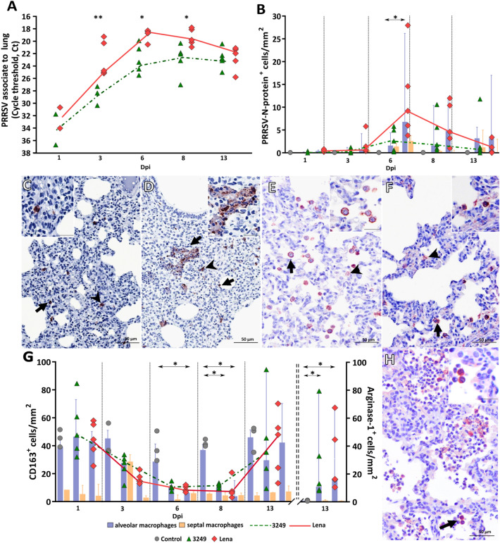
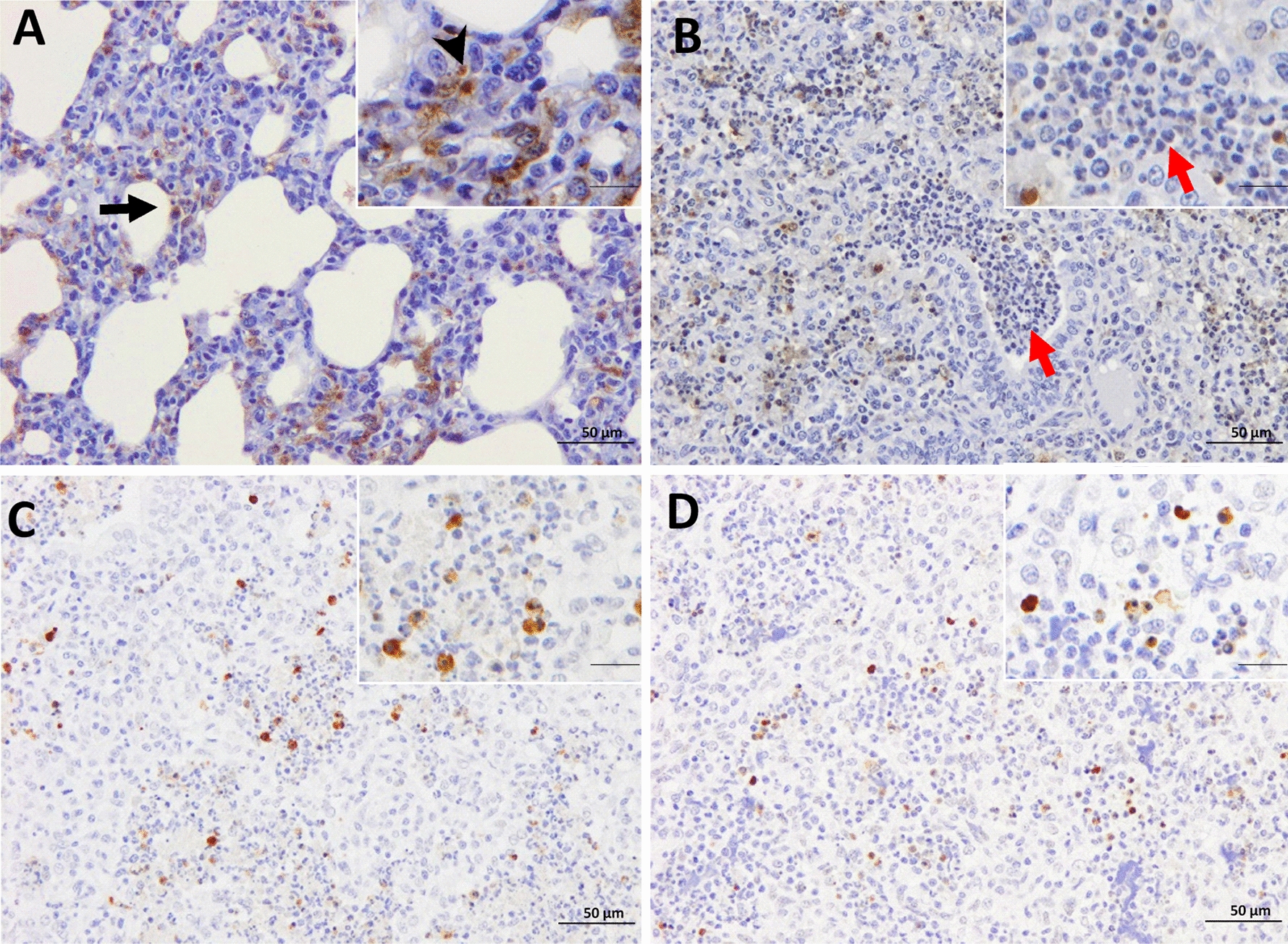
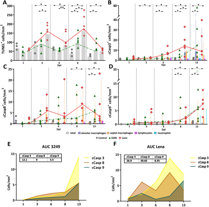
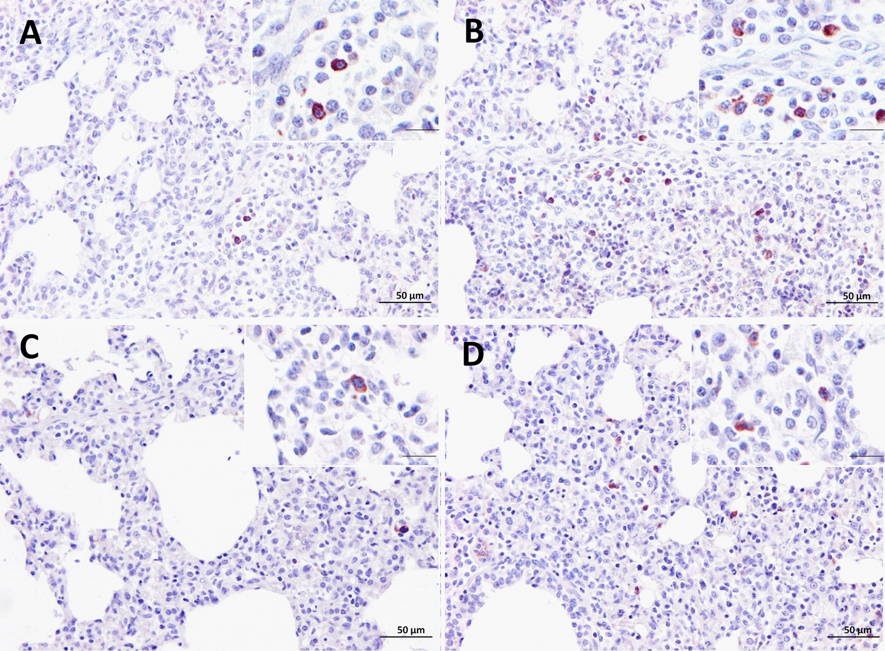
PRRSV-1 virulent strains cause high fever, marked respiratory disease and severe lesions in lung and lymphoid organs. Regulated cell death (RCD), such as apoptosis, necroptosis and pyroptosis, is triggered by the host to interrupt viral replication eliminating infected cells, however, although it seems to play a central role in the immunopathogenesis of PRRSV, there are significant gaps regarding their sequence and activation upon PRRSV-infection. The present study evaluated RCD events by means of caspases expression in the lung of PRRSV-1-infected pigs and their impact on pulmonary macrophage subpopulations and lung lesion. Conventional piglets were intranasally inoculated with the virulent subtype 3 Lena strain or the low virulent subtype 1 3249 strain and euthanised at 1, 3, 6, 8 and 13 dpi. Lena-infected piglets showed severe and early lung damage with a high frequency of PRRSV-N-protein+ cells, depletion of CD163+ cells and high viral load in the lung. The number of TUNEL+ cells was significantly higher than cCasp3+ cells in Lena-infected piglets during the first week post-infection. cCasp8 and to a lesser extent cCasp9 were activated by both PRRSV-1 strains after one week post-infection together with a replenishment of both CD163+ and Arg-1+ pulmonary macrophages. These results highlight the induction of other forms of RCD beyond apoptosis, such as, necroptosis and pyroptosis during the first week post-infection followed by the activation of, mainly, extrinsic apoptosis during the second week post-infection. The recovery of CD163+ macrophages at the end of the study represents an attempt to restore pulmonary macrophage subpopulations lost during the early stages of the infection but also a macrophage polarisation into M2 macrophages.

Keywords: PRRSV-1; cleaved caspase-8; lung; regulated cell death; virulent strain.


Conflict of interest statement

The authors declare that they have no competing interest.

Figures

Figure 1
Figure 1
Macroscopic and microscopic lung findings. Pictures show the macroscopic lung appearance A at necropsy of representative control (left), 3249 (middle) and Lena (right) animals euthanised at 13 dpi. Lungs from 3249 (middle) and Lena (right) infected pigs shown tan-mottled areas, atelectasis, rubbery consistency, but also consolidated areas are observed in cranial apical and middle lung lobes in Lena-infected pigs (right) (white arrows). Photomicrographs of the middle lung lobe B from control (left), 3249 (middle) and Lena (right) pigs euthanised at 13 dpi (H&E; bars 50 μm). Lung from 3249 (middle) and Lena (right) representative infected piglets show a moderate interstitial pneumonia characterised by thickening of the alveolar septa due to infiltrating lymphocytes and macrophages. Moreover, Lena-infected lung (right) exhibits extensive foci of suppurative bronchopneumonia with neutrophils, cellular debris, and multifocal chromatin clumps (arrow-head). Graphs display macroscopic C and microscopic D lung lesion score of each animal along the study. Bars indicate median values for each group.
Figure 2
Figure 2
PRRSV kinetics of viral load and immunohistochemical expression of PRRSV-N-protein, CD163 and Arg-1 in the lung. Graphs display the kinetics of viral load in the lung for Lena and 3249 strains. Viral load is represented by changes in the cycle threshold (Ct) A and the number of PRRSV-N-protein positive cells/mm2 B in the lung. Scatter dot plot shows individual values from each animal, bars and dashed lines indicate the median for each group and time points. Columns show the median with range of PAMs and septal macrophages (PIMs and interstitial macrophages). Photomicrographs of the middle lung lobe show the expression of PRRSV-N-protein in a representative 3249- C and Lena-infected pig D euthanised at 6 dpi (bar, 50 μm). Insets show in detail PRRSV-N-protein+ expression and viral particles (bar, 20 μm). Photomicrographs of the middle lung lobe show the expression of CD163 in a representative control E and Lena-infected F pig euthanised at 6 dpi (bar, 50 μm). Insets show in detail the immunolabelling of CD163 in the cell membrane and cytoplasm of PAMs (bar, 20 μm). Graph displays the number of CD163+ (primary axis) and Arg-1+ (secondary axis) cells/mm2 in the lung for each group and time point (G). Scatter dot plots show individual values from each animal, bars and dashed lines indicate the median for each group and time point. Columns represent the median with range of PAMs and septal macrophages. Photomicrograph of the middle lung lobe show the expression of Arg-1+ PAMs in a representative Lena-infected pig at 13 dpi H (bar, 50 μm). Inset shows in detail Arg-1 expression in PAMs (bar, 20 μm). Black arrows represent PAMs, and black arrow-heads interstitial macrophages.
Figure 3
Figure 3
Immunohistochemistry for TUNEL and cCasp3 in the lung. TUNEL expression in a representative 3249- A and Lena-infected pig B euthanised at 8 dpi (bar, 50 μm). Insets show in detail the immunolabelling of TUNEL in the nuclei and cytoplasm (bar, 20 μm). PAMs (black arrow), interstitial macrophages (black arrow-head) as well as TUNEL neutrophils (red arrows). cCasp3 expression in a representative 3249- C and Lena-infected pig D euthanised at 13 dpi (bar, 50 μm). Insets show in detail the expression of cCasp3 in the nuclei and cytoplasm of PAMs (bar, 20 μm).
Figure 4
Figure 4
Immunohistochemical expression of TUNEL, cCasp3, cCasp8 and cCasp9 in the lung along the experiment. Graph displays the number of TUNEL+ in the lung (cells/mm2) (A). Scatter dot plots show individual values from each animal and dashed lines indicate the median for each group and time points. Columns represent the median with range of total positive cells. Graph represents the number of cCasp3+ (B), cCasp8+ (C) cCasp9+ (D) in the lung (cells/mm2). Scatter dot plots show individual values from each animal and dashed lines indicate the median for each group and time point. Columns represent the median with range of PAMs, septal macrophages lymphocytes and neutrophils. One pig infected with 3249 strain at 6 dpi was considered an outlier (empty green triangle). Graph displays the AUC (median) for the frequency of cCasp3, cCasp8 and cCasp9 positive cells in 3249 E and Lena-infected F group.
Figure 5
Figure 5
Immunohistochemistry for cCasp8 and cCasp9 in lung tissue. cCasp8 expression in a representative 3249- A and Lena-infected pig B euthanised at 8 dpi (bar, 50 μm). Inset shows in detail the cCasp8 immunolabelling in the cytoplasm of interstitial macrophages (bar, 20 μm). cCasp9 expression in a representative 3249- C and Lena-infected pig D euthanised at 13 dpi (bar, 50 μm). Inset shows in detail the expression of cCasp9 in the cytoplasm of interstitial macrophages (bar, 20 μm).

References

    1. Morgan SB, Graham SP, Salguero FJ, Sánchez Cordón PJ, Mokhtar H, Rebel JMJ, Weesendorp E, Bodman-Smith KB, Steinbach F, Frossard JP. Increased pathogenicity of European porcine reproductive and respiratory syndrome virus is associated with enhanced adaptive responses and viral clearance. Vet Microbiol. 2013;163:13–22. doi: 10.1016/j.vetmic.2012.11.024. - DOI - PubMed
    1. Canelli E, Catella A, Borghetti P, Ferrari L, Ogno G, De Angelis E, Corradi A, Passeri B, Bertani V, Sandri G, Bonilauri P, Leung FC, Guazzetti S, Martelli P. Phenotypic characterization of a highly pathogenic Italian porcine reproductive and respiratory syndrome virus (PRRSV) type 1 subtype 1 isolate in experimentally infected pigs. Vet Microbiol. 2017;210:124–133. doi: 10.1016/j.vetmic.2017.09.002. - DOI - PubMed
    1. Karniychuk UU, Geldhof M, Vanhee M, Van Doorsselaere J, Saveleva TA, Nauwynck HJ. Pathogenesis and antigenic characterization of a new East European subtype 3 porcine reproductive and respiratory syndrome virus isolate. BMC Vet Res. 2010;6:30. doi: 10.1186/1746-6148-6-30. - DOI - PMC - PubMed
    1. Yuzhakov AG, Raev SA, Skrylev AN, Mishin AM, Grebennikova TV, Verkhovsky OA, Zaberezhny AD, Trus I, Nauwynck HJ, Aliper TI. Genetic and pathogenic characterization of a Russian subtype 2 PRRSV-1 isolate. Vet Microbiol. 2017;211:22–28. doi: 10.1016/j.vetmic.2017.09.017. - DOI - PubMed
    1. Sinn LJ, Klingler E, Lamp B, Brunthaler R, Weissenböck H, Rümenapf T, Ladinig A. Emergence of a virulent porcine reproductive and respiratory syndrome virus (PRRSV) 1 strain in Lower Austria. Porc Heal Manag. 2016;2:1–10. doi: 10.1186/s40813-016-0044-z. - DOI - PMC - PubMed

MeSH terms