Review
. 2021:647:173-208.
doi: 10.1016/bs.mie.2020.10.002. Epub 2020 Nov 18.

ER/K-link-Leveraging a native protein linker to probe dynamic cellular interactions

Affiliations
Review

ER/K-link-Leveraging a native protein linker to probe dynamic cellular interactions

Tejas M Gupte et al. Methods Enzymol. 2021.

Abstract

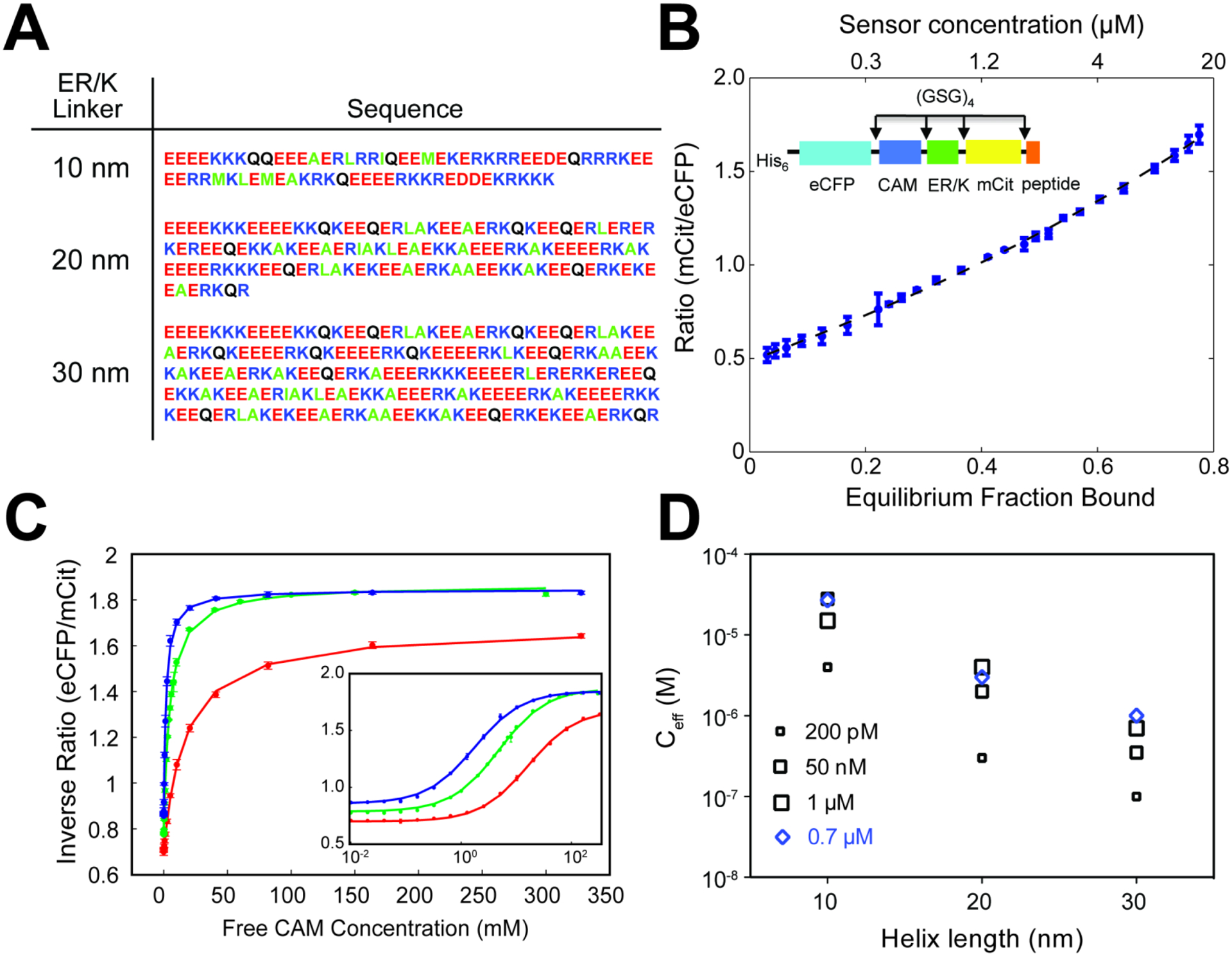
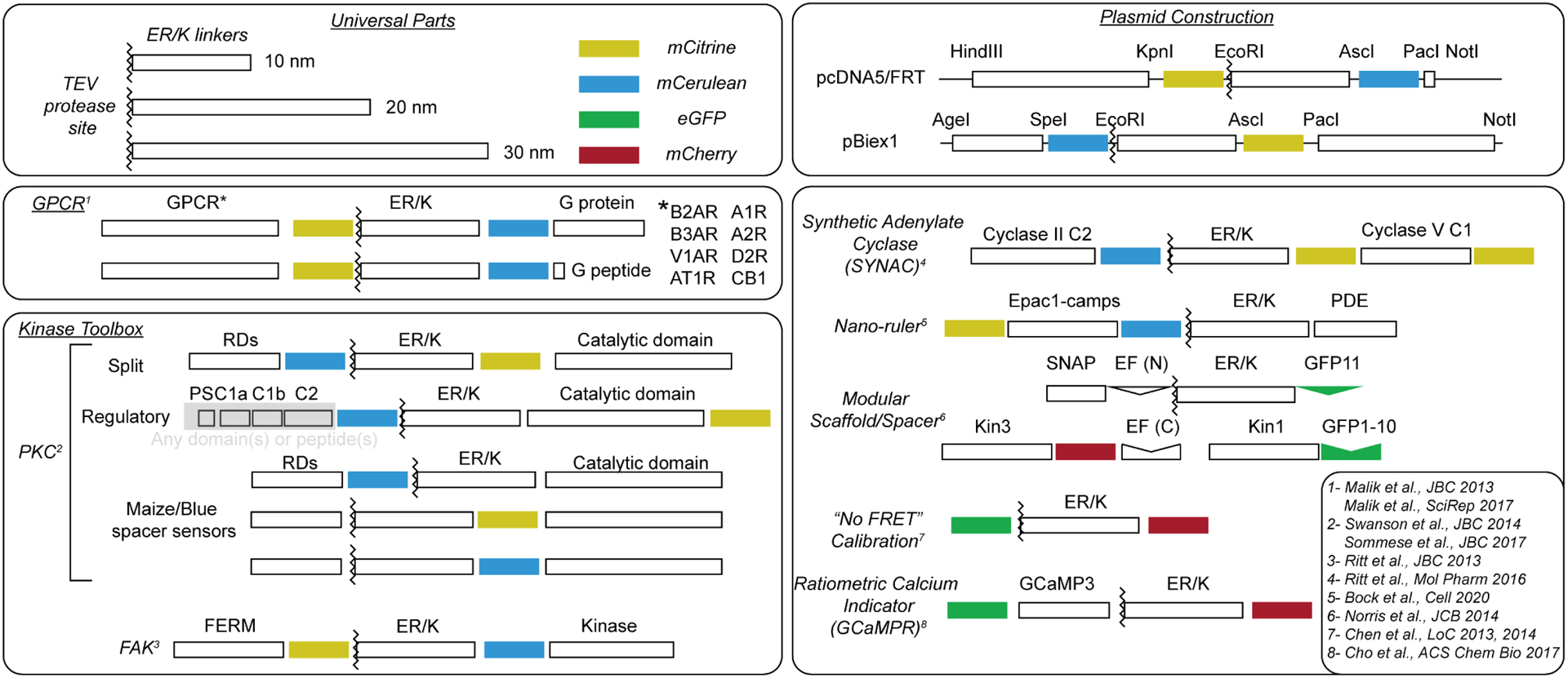
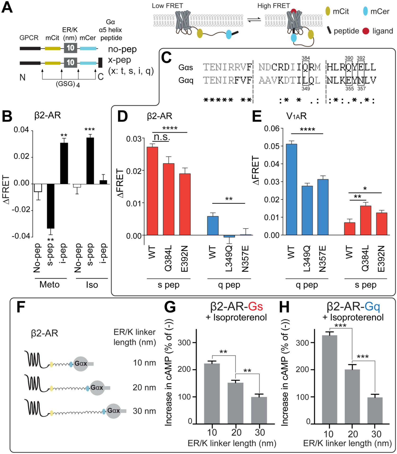
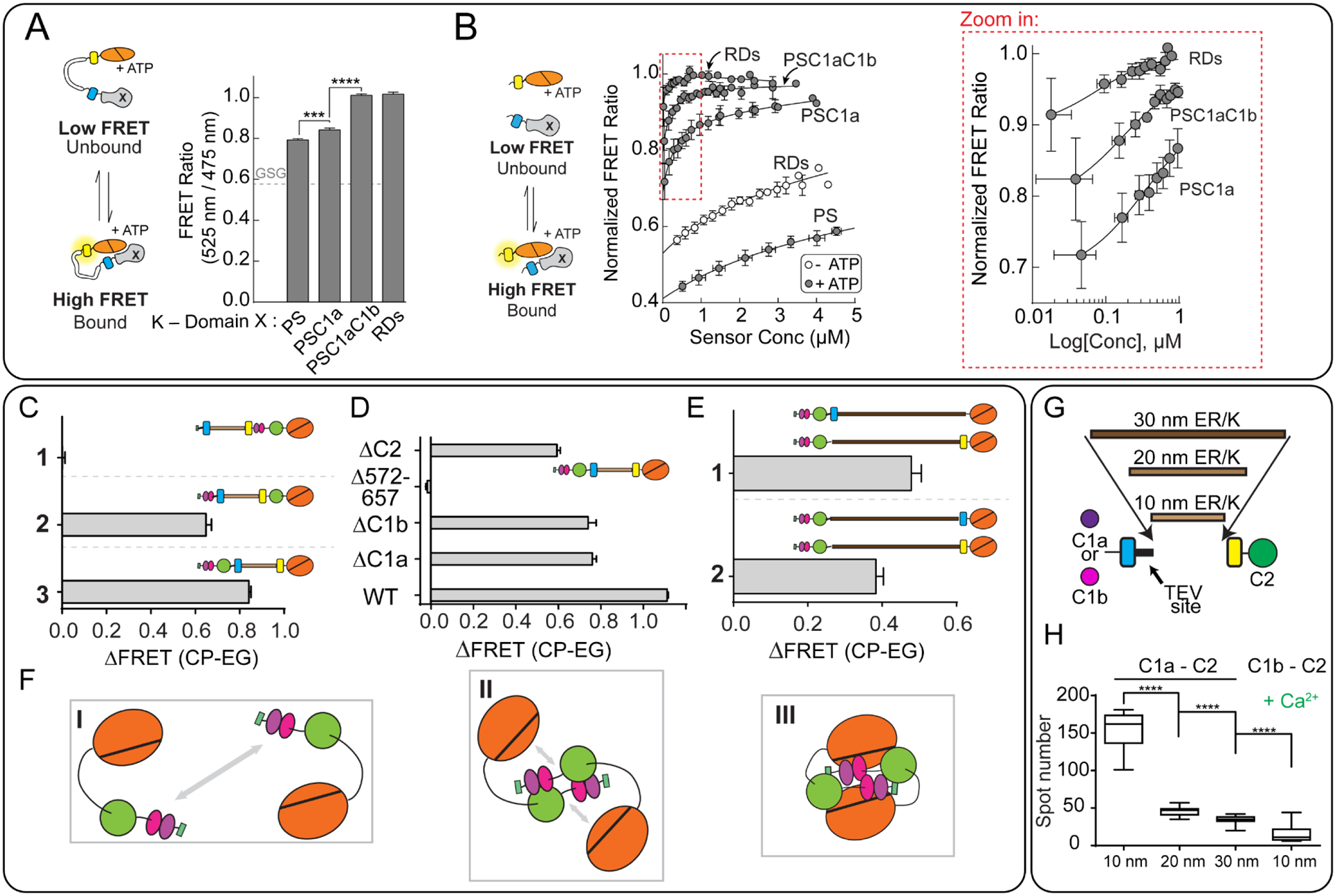
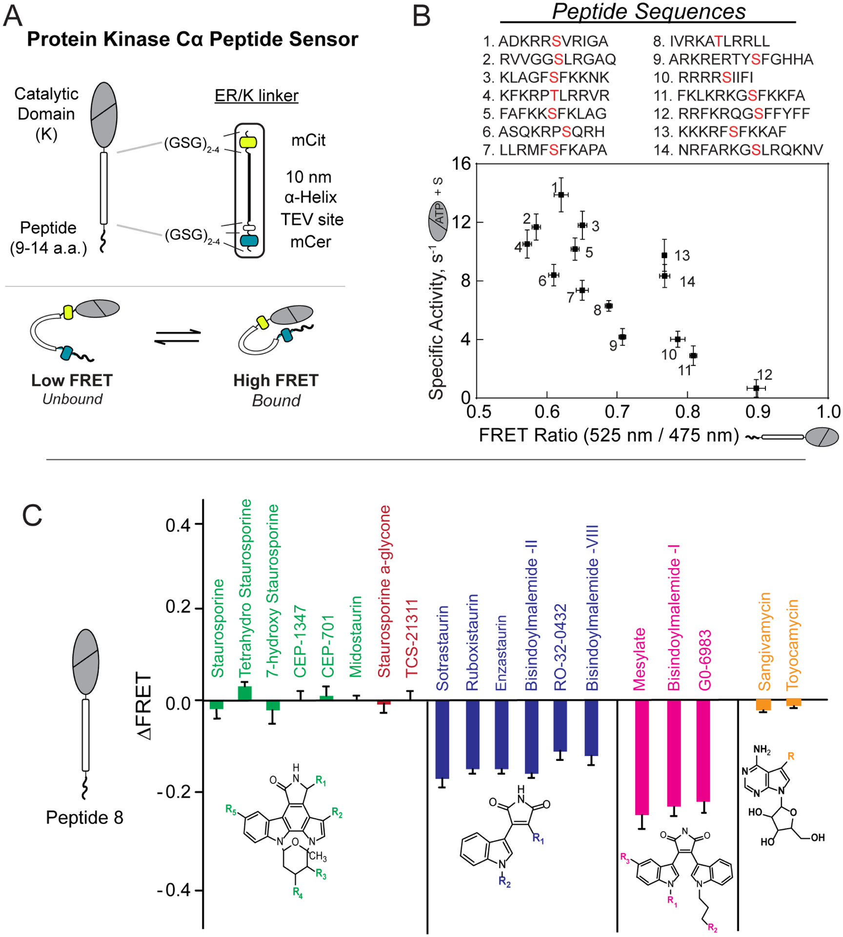
ER/K α-helices are a subset of single alpha helical domains, which exhibit unusual stability as isolated protein secondary structures. They adopt an elongated structural conformation, while regulating the frequency of interactions between proteins or polypeptides fused to their ends. Here we review recent advances on the structure, stability and function of ER/K α-helices as linkers (ER/K linkers) in native proteins. We describe methodological considerations in the molecular cloning, protein expression and measurement of interaction strengths, using sensors incorporating ER/K linkers. We highlight biological insights obtained over the last decade by leveraging distinct biophysical features of ER/K-linked sensors. We conclude with the outlook for the use of ER/K linkers in the selective modulation of dynamic cellular interactions.

Keywords: ER/K linker; FRET biosensors; GPCR; Kinases; Protein-protein interactions; Signaling; Single alpha helix.


Figures

Figure 1 –
Figure 1 –. ER/K linkers modulate the effective concentration of protein-protein interactions
A) Sequence of 10, 20 and 30 nm linkers used in ER/K-linked sensors. B) FRET ratio (mCit acceptor/eCFP donor) of ER/K-linked CaM-peptide sensor correlates linearly with fraction of sensors with proteins in the bound state. C, D) Effective concentrations (Ceff) engineered by 10, 20 and 30 nm ER/K linkers determined from (C) concentration-dependent quenching of FRET by unlabeled, free CaM protein. (D) Ceff for CaM interaction with peptides of indicated binding affinity (KD listed) and 10, 20 and 30 nm ER/K linker length. For all experiments: N = 4, and data are shown as mean ± SD. Panels A-D adapted from Sivaramakrishnan and Spudich, Systematic control of protein interaction using a modular ER/K α-helix linker. PNAS 2011. 108(51):20467–20472.
Figure 2-
Figure 2-. Schematics of sensors utilizing ER/K linkers
Generalized schematics of published sensor proteins that incorporate the ER/K linker. ER/K linkers have successfully been used to generate sensors employed to investigate cellular interactions involving GPCRs, kinases, motors, and second messenger signaling. ER/K-linked sensors are constructed so that the domains can be interchanged with relative ease during molecular cloning using restriction sites between protein elements (top right). Select citations for each sensor (superscript numbering) are also listed (bottom right).
Figure 3-
Figure 3-. GPCR biology investigated through the use of ER/K linkers.
A) Detailed schematic design of GPCR-Gα α5-helix peptide (Gα peptide) sensors (left). Cartoon rendering of GPCR-Gα peptide sensor interactions (right). B) Metoprolol stimulation of the indicated GPCR-Gα peptide sensors. Metoprolol (Meto), an inverse agonist of β1-AR signaling in the heart, suppresses the β2-AR-Gαs peptide interaction while stimulating interaction between β2-AR and Gαi peptide. In contrast, isoproterenol (Iso), a full agonist is shown to stimulate only the canonical β2-AR-Gαs peptide interaction. C) Sequence alignment of Gαs and Gαq α5-helix, from which the Gα peptides are derived. D,E) The indicated residues in panel C have been mutated to their counterpart residue from the other peptide. β2AR, is a Gαs coupled receptor (D) and shows diminished interaction with S-peptide mutated to look like Q-peptide. V1AR, a Gαq coupled receptor (E) shows enhanced interaction to S-peptide mutated to look like Q-peptide. F) Cartoon rendering of GPCR-Gα protein sensors with different length linkers. These protein sensors have the full-length G protein instead of the Gα α5-helix alone. G,H) β2-AR sensors tethered to either full length Gαs (G) or full length Gαq (H) with varying length ER/K linkers. Both sensors show decreases in cAMP production with increasing length of ER/K linkers. For all experiments: N ≥ 3, *, p≤0.05; **, p≤0.01; ***, p≤0.001; ****, p≤0.0001; n.s. not significant. Significance determined using Student’s t-test or ANOVA with Tukey’s post-hoc test where appropriate. Panels A (left) and B adapted from Malik et al., JBC 2013. This research was originally published in the Journal of Biological Chemistry. Detection of G Protein-selective G Protein-coupled Receptor (GPCR) Conformations in Live Cells. J Biol Chem. 2013; 288(24):17167–78. © the American Society for Biochemistry and Molecular Biology. Panels A(right), and C-E adapted from Semack et al., JBC 2016. This research was originally published in the Journal of Biological Chemistry. Structural elements in the Gαs and Gαq C-termini that mediate selective GPCR signaling. J Biol Chem. 2016; 19;291(34):17929–40. © the American Society for Biochemistry and Molecular Biology Panels F-H adapted from Gupte et al., Priming GPCR signaling through the synergistic effect of two G proteins. PNAS 2017. 114(14):3756–3761.
Figure 4 –
Figure 4 –. Investigating PKC structure and function using ER/K linkers
A) Cartoon diagram depicting interactions between regulatory domains and kinase domains when tethered with the ER/K linker (left). The observed FRET ratio for sensors with the kinase domain (K) and the indicated domains (right). PS – pseudosubtrate, C1a, C1b – diacylglycerol-sensitive regulatory domains, RDs – includes all regulatory domains (PS-C1a-C1b-C2). B) Cartoon diagram depicting bi-molecular interactions between the regulatory domains and kinase domain derived by proteolytic cleavage of a TEV-protease site at the N-terminus of the ER/K linker (left). FRET ratio of the indicated pairings between regulatory domains and kinase domain (center). The region highlighted by the red box is depicted in the zoom-in on the right. C-E) Change in FRET ratio in the indicated PKC sensors (ΔFRET) following activation. CP (calcium and PMA stimulation); EG (buffer containing EGTA). C) By inserting the ER/K linker at various points in the protein, the domains that participate in the interaction can be discerned. 1- Sensor with pseudo-substrate separated from the regulatory domains. 2- pseudo-substrate and C1a/b separated from the C2 and kinase domains. 3- regulatory domains separated from the kinase domain. D) PKC sensors with various truncations. The changes in ΔFRET following PKC activation indicated the relative importance of each excluded domain to the interaction. For instance, residues 572–657 that constitute the C-terminus of the kinase domain are essential to stimulate interactions upon PKC activation. E) The change in FRET ratio for a pair of PKC sensors, each containing 30 nm linkers, and either an mCitrine or mCerulean fluorophore. By locating the fluorophores at opposite ends of the ER/K linker (1), a greater change in FRET is observed upon calcium and PMA stimulation than when they are located on the same side of the linker (2), indicating that the interaction between molecules likely involves close interaction between the regulatory and kinase domains on at least two separate proteins. F) A proposed model for the interactions probed by the experiments in panels C-E G) Diagram of the sensors used in panel H. These sensors contain either the C1a or C1b domains linked to C2 with varying length ER/K linkers. H) Decrease in observed spots in a microscopy assay using the indicated sensors depicted in (G). C1a, but not C1b aggregates in response to calcium stimulation in a manner proportional to the length of the ER/K linker. For all experiments: N ≥ 3, **, p≤0.01; ****, p≤0.0001. Significance determined using ANOVA with Tukey’s post-hoc test. Panels A and B adapted from Sommese et al., JBC 2017 This research was originally published in the Journal of Biological Chemistry. The role of regulatory domains in maintaining auto-inhibition in the multi-domain kinase PKCα. J Biol Chem. 2017; 292(7):2873–2880. © the American Society for Biochemistry and Molecular Biology Panels C-F adapted from Swanson et al., JBC 2014 This research was originally published in the Journal of Biological Chemistry. Conserved modular domains team up to latch-open Active protein kinase Cα. J Biol Chem. 2014; 20;289(25):17812–29. © the American Society for Biochemistry and Molecular Biology Panels G and H adapted from Swanson et al., PLoSone 2016
Figure 5 –
Figure 5 –. Substrate peptide-kinase interactions and their allosteric modulation by small molecules probed using ER/K linkers
A) Cartoon schematic of PKC substrate peptide biosensor (top) and mechanism of action (bottom) B) Correlation between kinase activity and FRET ratio observed for 14 different peptides derived from phosphorylated substrates of PKC (phosphorylated Ser/Thr residue is highlighted in red). Lower FRET ratios (affinities) for peptides were observed to correlate with higher activity. C) Families of structurally similar small molecule kinase inhibitors led to similar observed changes in FRET ratio in PKC biosensors. Small molecules with a purported bitopic mode of binding (navy, magenta) were more effective in disrupting the kinase/substrate interaction. For all experiments: N ≥ 3, and data are shown as mean ± SE. Panels A and B adapted from Sommese et al., JBC 2016 This research was originally published in the Journal of Biological Chemistry. Substrate Affinity Differentially Influences Protein Kinase C Regulation and Inhibitor Potency. J Biol Chem. 2016; 291(42):21963–21970. © the American Society for Biochemistry and Molecular Biology Panel C adapted with permission from (Ma et al., Biochemistry 2018). Copyright (2018) American Chemical Society

Similar articles

Cited by

References

    1. Al-Osta I, Mucha M, Pereda D, Piqué-Gili M, Okorocha AE, Thomas R, et al. Imaging Calcium in Hippocampal Presynaptic Terminals With a Ratiometric Calcium Sensor in a Novel Transgenic Mouse. Front Cell Neurosci 2018;12:209. 10.3389/fncel.2018.00209. - DOI - PMC - PubMed
    1. Antal CE, Callender JA, Kornev AP, Taylor SS, Newton AC. Intramolecular C2 Domain-Mediated Autoinhibition of Protein Kinase C βII. Cell Rep 2015;12:1252–60. 10.1016/j.celrep.2015.07.039. - DOI - PMC - PubMed
    1. Batchelor M, Wolny M, Baker EG, Paci E, Kalverda AP, Peckham M. Dynamic ion pair behavior stabilizes single -helices in proteins. J Biol Chem 2019;294:3219–34. 10.1074/jbc.RA118.006752. - DOI - PMC - PubMed
    1. Bierzynski A, Kim PS, Baldwin RL. A salt bridge stabilizes the helix formed by isolated C-peptide of RNase A. Proc Natl Acad Sci U S A 1982;79:2470–4. 10.1073/pnas.79.8.2470. - DOI - PMC - PubMed
    1. Bock A, Annibale P, Konrad C, Hannawacker A, Anton SE, Maiellaro I, et al. Optical Mapping of cAMP Signaling at the Nanometer Scale. Cell 2020;182:1–12. 10.1016/j.cell.2020.07.035. - DOI - PMC - PubMed

Publication types